Understanding Kidney Function and Urinary Tract Disorders
This content explains the sophisticated regulatory functions of the kidneys, including filtration, waste removal, electrolyte balance, and endocrine roles. It details urinary tract obstruction, obstructive uropathy, and the pathophysiology, composition, and clinical manifestations of kidney stones (urolithiasis).
The kidneys are sophisticated regulatory organs that maintain the body's internal equilibrium. Beyond simply producing urine, they serve as vital filtration units that manage blood composition, fluid volume, and systemic hormonal balance. When these processes are disrupted—whether by physical blockages, crystal formation, or inflammatory disease—the resulting alterations can lead to severe systemic complications, ranging from localized pain to life-threatening renal failure.
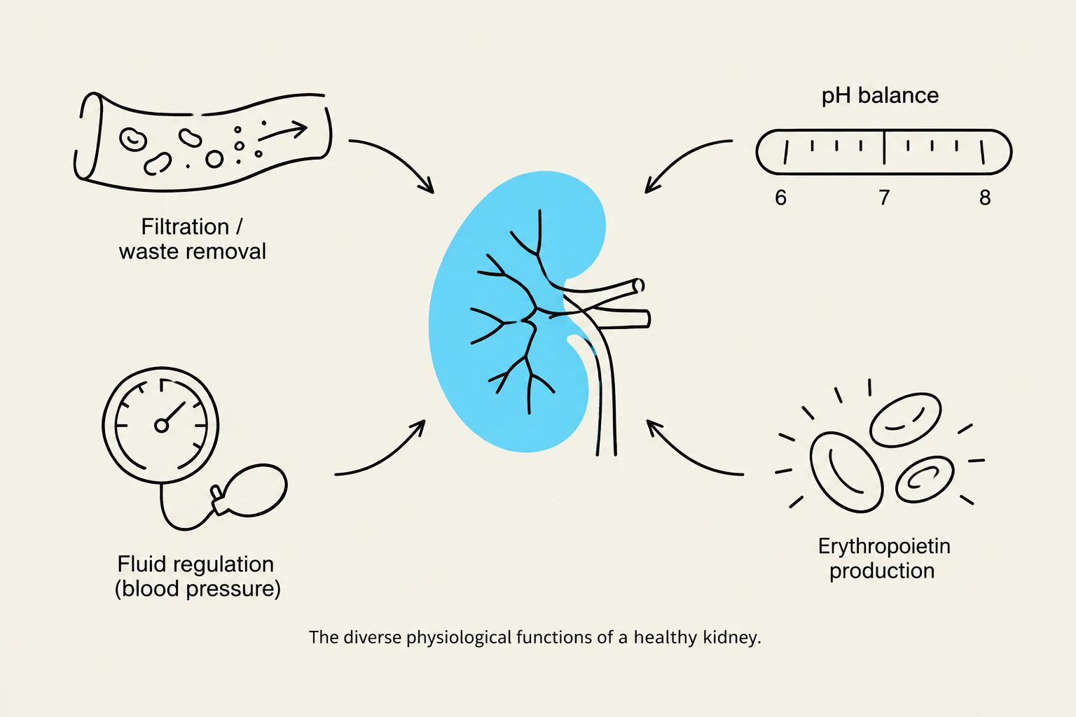
The diverse physiological functions of a healthy kidney, including filtration, fluid regulation, pH balance, and erythropoietin production.
The Physiological Role of the Kidneys
To understand renal pathology, one must first appreciate the diverse functions of a healthy kidney. The kidneys are responsible for:
Filtration and Waste Removal: Clearing metabolic waste products from the blood.
Electrolyte and Acid/Base Balance: Maintaining precise concentrations of ions and regulating blood pH.
Fluid and Pressure Regulation: Controlling blood volume and blood pressure.
Endocrine Functions: Activating Vitamin D (essential for bone health) and producing erythropoietin, the hormone that stimulates red blood cell production.
Urinary Tract Obstruction and Obstructive Uropathy
An obstruction occurs when an anatomic or functional defect blocks the flow of urine. This blockage leads to obstructive uropathy, which refers to the anatomic changes in the urinary system caused by the obstruction. The clinical severity of an obstruction is determined by its location, whether it is complete or partial, its duration, and whether it affects one or both upper urinary tracts.
Complications of Upper Tract Obstruction
Diagram illustrating normal urinary tract anatomy and the structural changes associated with hydronephrosis, hydroreter, and ureterohydronephrosis due to obstruction.
When the obstruction occurs in the upper urinary tract (the kidneys or ureters), several structural changes may occur:
Hydroureter: Dilation of the ureters.
Hydronephrosis: Dilation of the renal pelvis and calyces.
Ureterohydronephrosis: A combination where both the ureters and the renal pelvis/calyces are dilated.
Tubulointerstitial Fibrosis: The deposition of excessive extracellular matrix, which leads to the destruction and death of nephrons.
Physiological Responses to Obstruction
The body attempts to adapt to the loss of renal function. If one kidney is lost to obstructive disease, the unaffected kidney may undergo compensatory hypertrophy (an increase in size) and hyperfunction to maintain homeostasis.
Once an obstruction is relieved, patients often experience post-obstructive diuresis. This is a physiological response characterized by a large increase in urine output, which can lead to dehydration and electrolyte imbalances due to alterations in how the tubules transport water and solutes.
Kidney Stones (Urolithiasis)
Kidney stones, also known as renal calculi or urolithiasis, are masses composed of crystals, proteins, or mineral salts that form within the urinary tract.
Pathophysiology of Stone Formation
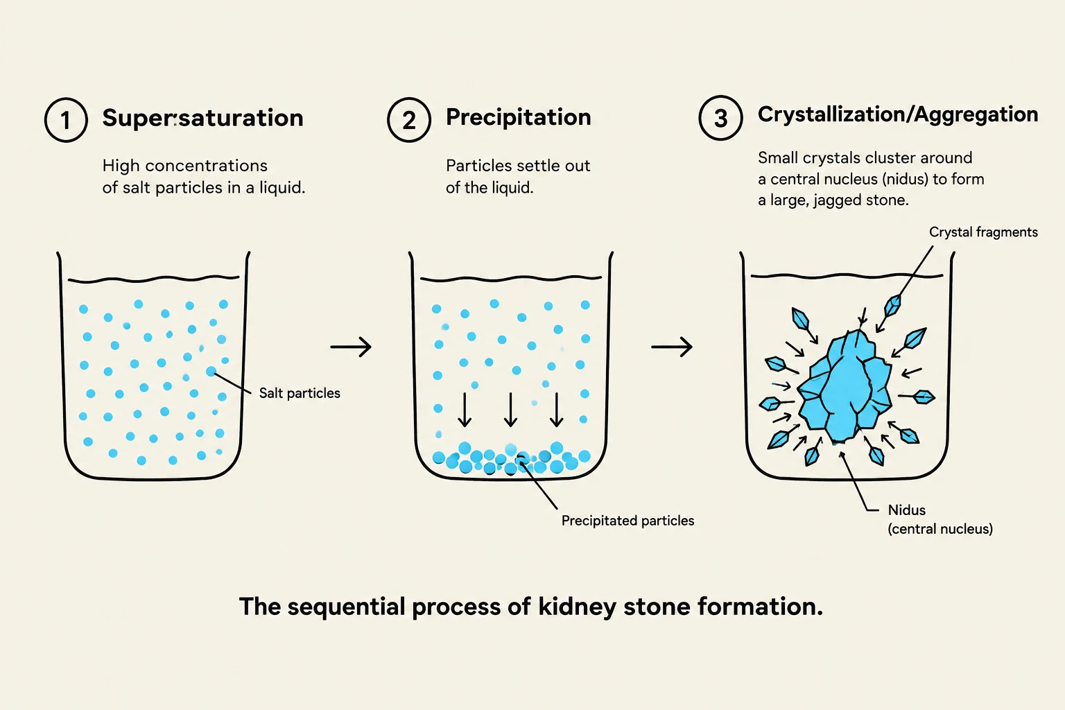
The sequential process of kidney stone formation, illustrating supersaturation, precipitation, and crystallization/aggregation.
The formation of a stone follows a specific sequence of events:
Supersaturation: This occurs when the concentration of salts in the urine exceeds the volume's ability to keep them dissolved.
Precipitation: The salts transition from a liquid state to a solid state.
Crystallization or Aggregation: Crystals grow from a small nucleus (nidus) into larger stones by embedding in a matrix.
Common Stone Compositions
The type of stone is often determined by urine pH and metabolic factors:
Calcium Oxalate and Calcium Phosphate: The most common types, accounting for 70%–80% of cases.
Struvite: Composed of magnesium, ammonium, and phosphate; these typically form in alkaline urine during infections (e.g., Klebsiella or Pseudomonas).
Uric Acid: Often associated with gout or genetic disorders of amino acid metabolism.
Cystine/Xanthine: Formed in the presence of low urine pH.
Staghorn Calculi: Massive stones that fill the minor and major calyces of the kidney.
Clinical Tip: The Role of pH
Urine pH is a critical determinant in stone formation. Alkaline urine increases the risk of calcium phosphate stones, while acidic urine increases the risk of uric acid stones. Inhibitors such as potassium citrate, pyrophosphate, and magnesium can help prevent stone formation.
Clinical Manifestations
Patients presenting with kidney stones often exhibit:
Renal Colic: Moderate to severe pain originating in the flank and radiating to the groin (indicating obstruction of the renal pelvis or proximal ureter).
Hematuria: The presence of blood in the urine.
Gastrointestinal Symptoms: Nausea and vomiting may accompany severe pain.
Risk Factors and Demographics
While anyone can develop urolithiasis, certain factors increase susceptibility:
Gender: More common in males.
Age: Most cases develop before age 50.
Fluid Intake: Inadequate fluid intake is the most prevalent risk factor.
Environment: Geographic location, temperature, and humidity can influence risk.
Summary Table: Stone Composition and Risk Factors
Stone Type
Composition
Key Risk Factor/Context
Calcium-based
Calcium oxalate/phosphate
Most common (70-80%); Alkaline pH increases risk
Struvite
Mg, Ammonium, Phosphate
Alkaline urine; Infections
Uric Acid
Uric acid
Acidic urine; Gout; Amino acid disorders
Cystine
Cystine
Low urine pH
Keep reading for free.
Create an account to finish this reading and make it part of your study routine.