This content explains the body's inflammatory response to injury and infection, detailing acute vs. chronic inflammation, vascular and cellular mechanisms, the role of leukocytes, common pathogens, and potential clinical complications like sepsis and autoimmune diseases.
When the body encounters a threat—whether it is a physical injury, a toxin, or a invading microorganism—it does not remain passive. Instead, it initiates a complex, highly coordinated biological response known as inflammation. While often perceived as a negative symptom (like swelling or pain), inflammation is actually a vital protective mechanism designed to eliminate the cause of injury, clear out necrotic (dead) cells, and initiate the repair process. However, the line between a helpful response and a harmful one is thin; if the response is excessive or fails to resolve, it can lead to significant tissue damage or systemic illness.
The Mechanisms of Inflammation
Inflammation is categorized based on its duration and the specific cellular players involved. Understanding these distinctions is crucial for clinical diagnosis.
Acute vs. Chronic Inflammation
Acute inflammation is the body's immediate response to injury. It is characterized by a rapid onset and a relatively short duration. Clinically, this is often recognized by the "cardinal signs": redness, heat, swelling, and pain. The primary cellular driver in this stage is the neutrophil, which arrives quickly to fight pathogens and clean up debris.
In contrast, chronic inflammation is a prolonged response that can persist for months or even years. This occurs when the stimulus cannot be eliminated or when the immune system mistakenly targets healthy tissue. Unlike the neutrophil-heavy acute phase, chronic inflammation is characterized by the presence of macrophages and lymphocytes. While intended to resolve the issue, chronic inflammation often leads to tissue destruction and fibrosis (the formation of scar tissue).
The Vascular and Cellular Response
The inflammatory process follows a predictable sequence of vascular and cellular changes to ensure immune cells reach the site of injury.
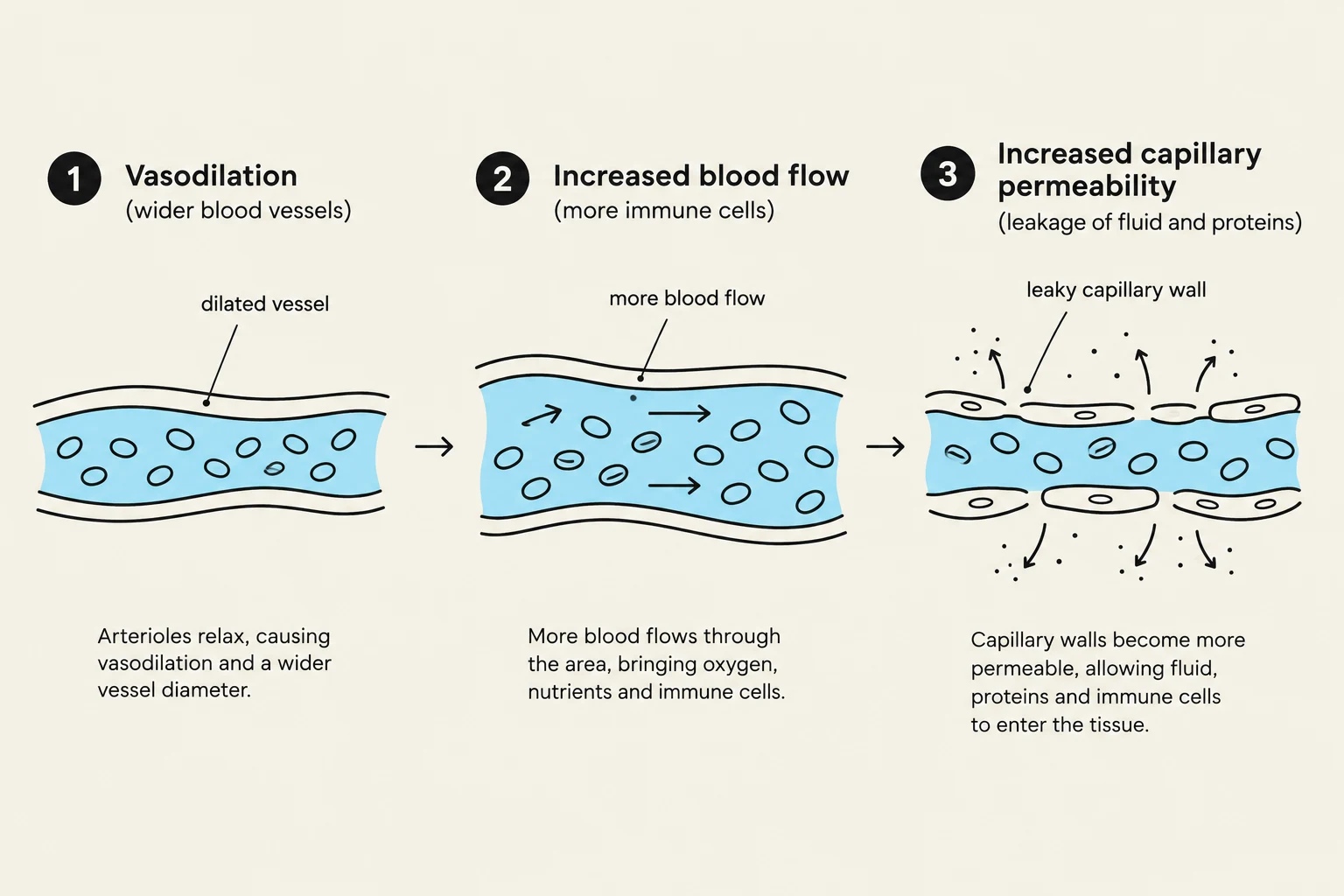
Vascular Changes: Upon injury, chemical mediators trigger vasodilation, which increases blood flow to the area (causing redness and heat). Simultaneously, capillary permeability increases, allowing plasma and immune cells to leak out of the bloodstream and into the interstitial space. This accumulation of fluid in the tissue results in edema (swelling).
Cellular Response and Chemotaxis: To guide immune cells to the exact location of the threat, the body uses chemical signals called cytokines and chemokines. Through a process called chemotaxis, leukocytes (white blood cells) follow these chemical gradients to migrate toward the injury site.
Phagocytosis: Once at the site, specialized cells perform phagocytosis—the process of engulfing and destroying foreign invaders, bacteria, or dead cellular debris.
This diagram illustrates the three stages of inflammation: vasodilation, increased blood flow, and increased capillary permeability, showing how arterioles relax, blood flow increases, and capillaries become more permeable to allow fluid, proteins, and immune cells to enter the tissue.
The Role of Leukocytes
Leukocytes, or white blood cells, are the "soldiers" of the immune system. Each type has a specialized role in the inflammatory and infectious response.
Neutrophils: The first responders of the acute inflammatory phase. They are highly efficient at phagocytosis and are often the first cells to arrive at a site of bacterial infection.
Monocytes and Macrophages: Monocytes circulate in the blood; once they migrate into the tissues, they differentiate into macrophages. Macrophages are "big eaters" that play a central role in both acute and chronic inflammation, acting as long-term scavengers and orchestrating the transition to tissue repair.
Lymphocytes: These are the primary cells of the adaptive immune response. They are central to chronic inflammation and are responsible for recognizing specific pathogens and creating immunological memory.
Eosinophils: These cells are specialized for fighting parasitic infections and are also heavily involved in allergic reactions.
Basophils and Mast Cells: These cells are key players in allergic responses and inflammation. Mast cells, located in tissues, release histamine and other mediators that drive the vascular changes (vasodilation and permeability) seen in early inflammation.
Exam Tip: If a clinical case describes a patient with a sudden onset of swelling and high neutrophil counts, think Acute Inflammation. If the case describes long-term tissue scarring and high lymphocyte/macrophage counts, think Chronic Inflammation.
Infection and Pathogens
An infection occurs when pathogenic microorganisms successfully invade the body and begin to multiply. The nature of the inflammatory response is often dictated by the type of pathogen involved.
Common Pathogens
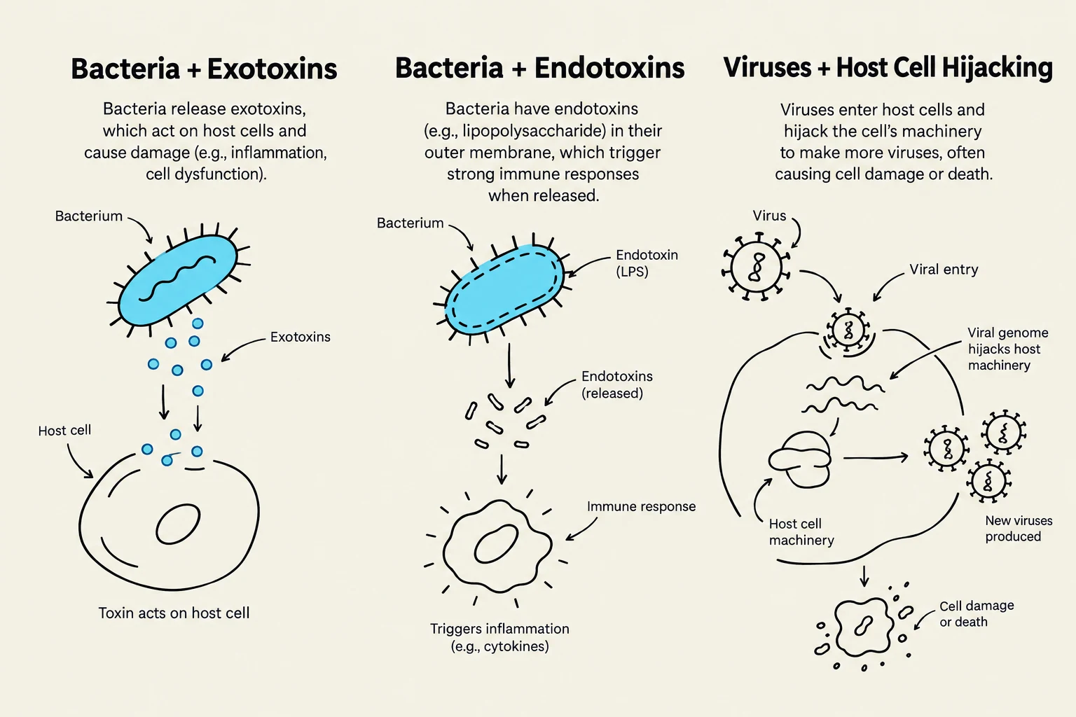
Bacteria: These can be highly virulent. Some bacteria secrete exotoxins (toxins released by live bacteria) to damage host cells, while others possess endotoxins (such as Lipopolysaccharide/LPS in Gram-negative bacteria) which are part of their cell wall and can trigger massive inflammatory responses when the bacteria die.
Viruses: These are obligate intracellular parasites. They must enter a host cell to use its machinery for replication, which often results in the death (lysis) of the host cell.
Fungi and Parasites: These can cause both localized and systemic infections, often triggering intense, specialized inflammatory responses (such as the eosinophil response in parasitic infections).
This illustration depicts three ways pathogens can affect host cells: bacteria releasing exotoxins, bacteria releasing endotoxins, and viruses hijacking host cell machinery to replicate.
Clinical Complications
While inflammation is a survival mechanism, it can become a pathological force if it loses regulation.
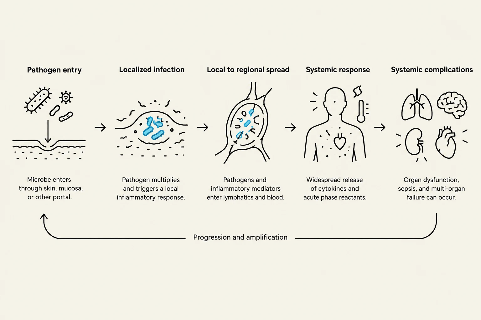
One of the most severe complications is sepsis, a life-threatening systemic inflammatory response to an infection. In sepsis, the inflammatory mediators enter the bloodstream and trigger widespread damage to organs throughout the body. Another complication is the development of autoimmune diseases, where the immune system's ability to distinguish "self" from "non-self" fails, leading it to attack the body's own healthy tissues. Finally, as noted previously, uncontrolled chronic inflammation can lead to permanent tissue destruction and loss of organ function through excessive fibrosis.
This diagram illustrates the progression of an infection from initial pathogen entry to localized infection, regional spread, systemic response, and potential systemic complications such as organ dysfunction.
Keep reading for free.
Create an account to finish this reading and make it part of your study routine.