Understanding Hypersensitivity Reactions in the Immune System
This content explains the four main types of hypersensitivity reactions (Type I, II, III, and IV) in the immune system, detailing their mechanisms, time courses, and common examples.
The immune system is a sophisticated defense network designed to protect the body from pathogens and abnormal cells. However, this system does not always distinguish perfectly between dangerous invaders and harmless environmental substances. When the immune system mounts an aggressive response against a perceived, but otherwise harmless, threat, the result is tissue damage. These reactions are known as hypersensitivities.
Hypersensitivities are not all the same; they are categorized into different types based on two primary factors: their time course (how quickly they appear) and the specific components of the immune system involved (whether they are mediated by antibodies or T cells).
Immediate Hypersensitivity (Type I)
Often referred to as acute hypersensitivity or common allergies, Type I reactions are characterized by their incredibly rapid onset. These reactions can begin within seconds of contact with an allergen—an antigen that triggers an allergic reaction.
The Mechanism of Sensitization and Reaction
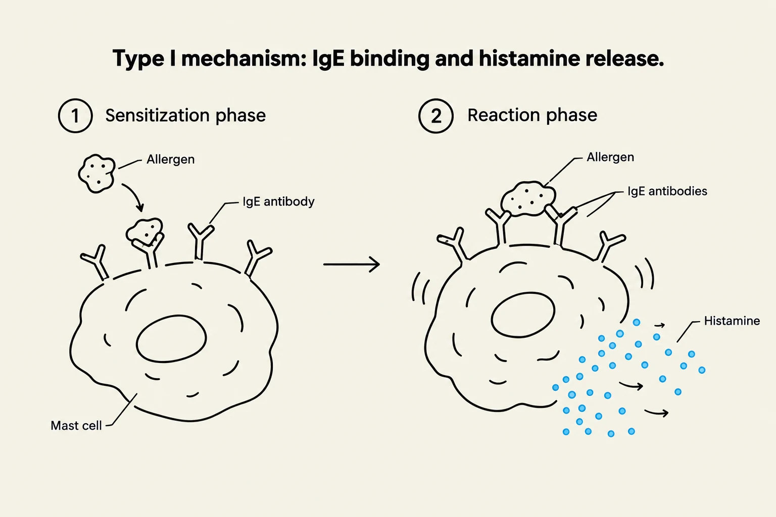
Diagram illustrating the Type I hypersensitivity mechanism involving IgE binding to allergens and subsequent histamine release from mast cells.
The process begins with an initial encounter that is typically asymptomatic. During this first contact, the body becomes sensitized to the allergen. The immune system produces specific IgE antibodies that target the allergen. These IgE antibodies then bind to the surfaces of mast cells and basophils.
When the individual encounters the same allergen again, the allergen binds to the IgE already sitting on these cells. This triggers a massive, sudden histamine release. Histamine is a potent inflammatory chemical that causes vasodilation (widening of blood vessels) and increased capillary permeability (making vessels "leaky").
Local vs. Systemic Reactions
Depending on where the allergen enters the body, the symptoms can vary significantly:
Local Reactions: These involve mast cells located in the skin or the mucosal linings of the respiratory or gastrointestinal tracts. Common symptoms include a runny nose, itchy hives, watery eyes, or asthma if the allergen is inhaled.
Systemic Reactions: If the allergen enters the bloodstream (such as through a bee sting), it can trigger a body-wide response known as anaphylactic shock.
Clinical Alert: Anaphylactic Shock
Anaphylactic shock is a life-threatening emergency. It is characterized by the constriction of bronchioles (making breathing difficult) and extreme vasodilation, which leads to a dangerous drop in blood volume and potential circulatory collapse. The standard emergency treatment is the administration of epinephrine.
Subacute Hypersensitivities (Types II and III)
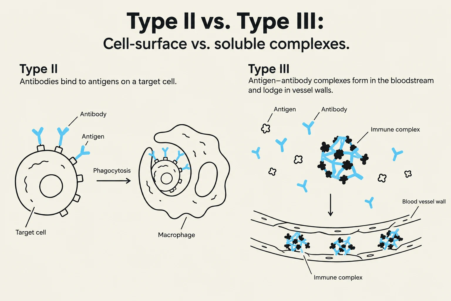
Diagram illustrating Type II hypersensitivity where antibodies bind to antigens on a target cell leading to phagocytosis, and Type III hypersensitivity where antigen-antibody complexes form in the bloodstream and lodge in blood vessel walls.
Subacute hypersensitivities are mediated by antibodies—specifically IgM and IgG. Unlike the immediate reactions of Type I, these have a slower onset, typically taking 1–3 hours to manifest, and a longer duration, lasting between 10–15 hours.
Cytotoxic Hypersensitivity (Type II)
In a Type II reaction, antibodies bind directly to antigens located on the surface of specific body cells. Once bound, these antibodies trigger the destruction of the cell through two main pathways: phagocytosis (where cells like macrophages eat the target) or complement-mediated lysis (where the complement system creates holes in the cell membrane). A classic clinical example of this is a mismatched blood transfusion reaction.
Immune Complex Hypersensitivity (Type III)
Type III reactions occur when antigens are widely distributed throughout the blood or body. When these widely dispersed antigens meet antibodies, they form insoluble antigen-antibody complexes.
Because these complexes are insoluble, they cannot be easily cleared from the system. Instead, they settle in specific tissues, such as blood vessel walls or kidney tissues. Once lodged, they trigger intense inflammation, which leads to local cell lysis and the recruitment of neutrophils that kill the surrounding tissue. A well-known example of a systemic Type III hypersensitivity is systemic lupus erythematosus (SLE).
Delayed Hypersensitivity (Type IV)
The final category, Type IV hypersensitivity, is unique because it is not mediated by antibodies at all. Instead, it is a T cell-mediated response. Because it relies on the activation and migration of cells rather than the immediate release of pre-formed chemicals, it has a slow onset, often taking 1–3 days to appear.
The Cellular Mechanism
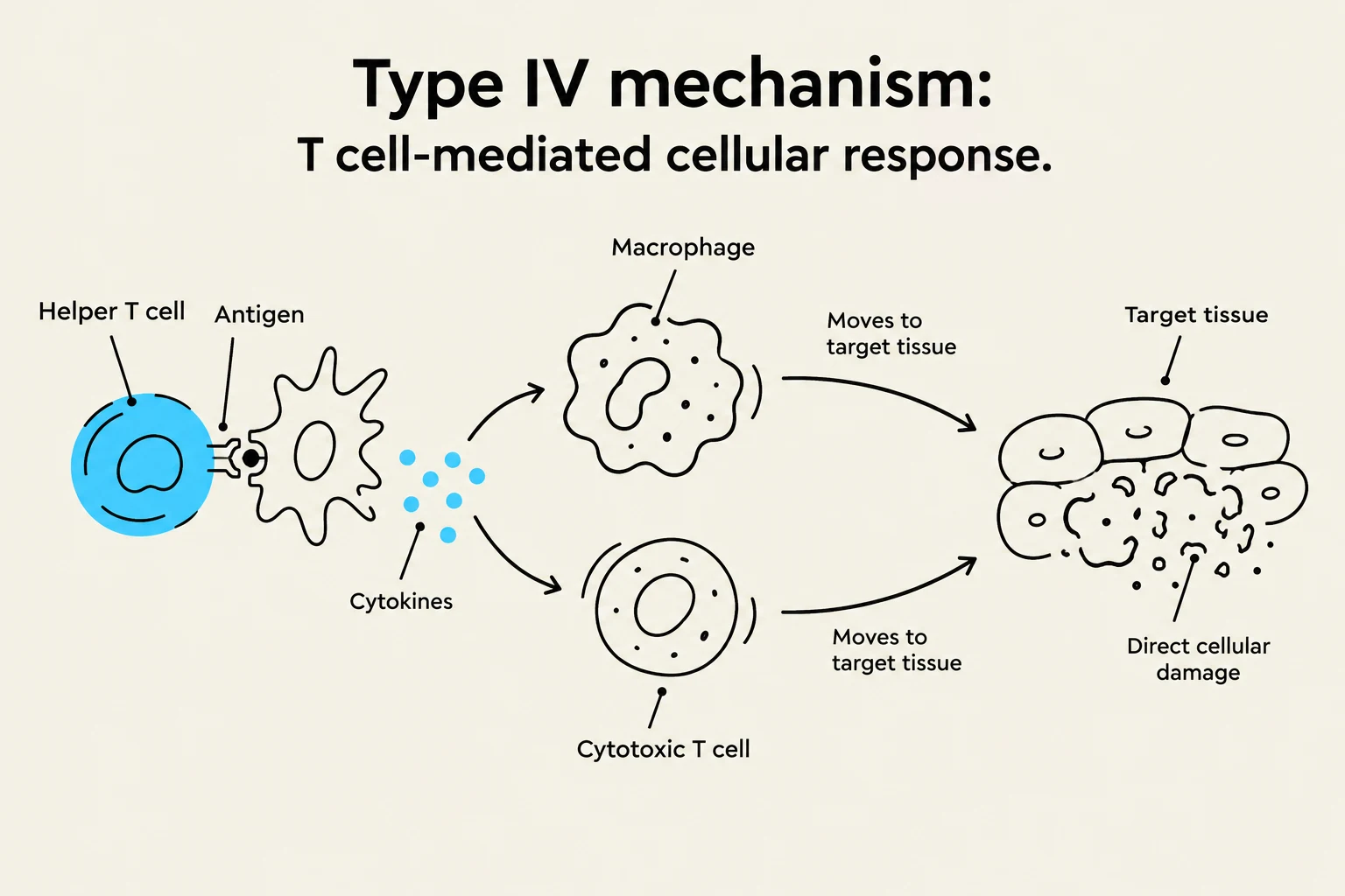
Diagram illustrating the Type IV mechanism, a T cell-mediated cellular response involving helper T cells, antigens, macrophages, cytokines, cytotoxic T cells, and target tissue leading to direct cellular damage.
The mechanism of Type IV hypersensitivity involves a complex interaction between different lymphocyte populations:
Helper T cells recognize the antigen and release cytokines.
These cytokines activate macrophages and cytotoxic T cells.
The activated macrophages and cytotoxic T cells then move into the tissue to cause direct cellular damage.
Haptens and Clinical Examples
In many cases, the trigger for a Type IV reaction is a hapten. Haptens are small molecules (such as those found in certain chemicals or plant toxins) that are not antigenic on their own but become antigenic when they bind to larger body proteins.
Common examples include:
Allergic contact dermatitis: Such as the skin irritation caused by touching poison ivy.
The TB skin test: This medical test relies on the delayed T cell response to detect previous exposure to the bacteria that causes tuberculosis.
Summary Table of Hypersensitivities
Type
Name
Mediator
Time Course
Example
Type I
Immediate / Acute
IgE (Mast cells/Basophils)
Seconds to minutes
Allergies, Anaphylaxis
Type II
Cytotoxic
IgG / IgM
1–3 hours
Mismatched blood transfusion
Type III
Immune Complex
IgG / IgM (Complexes)
1–3 hours
Systemic Lupus Erythematosus (SLE)
Type IV
Delayed
T cells (Helper & Cytotoxic)
1–3 days
Poison ivy, TB skin test
Keep reading for free.
Create an account to finish this reading and make it part of your study routine.