Understanding Homeostasis: Fluids, Electrolytes, and Acid-Base Balance
This guide explains the fundamental concepts of homeostasis in the human body, focusing on the critical balance of fluids, electrolytes, and pH. It details the roles of various body systems, common imbalances like hypervolemia and hypovolemia, and the interpretation of arterial blood gases (ABGs).
In the complex environment of the human body, survival depends on homeostasis—the ability to maintain stable internal conditions despite constant changes in the external environment. For cells to work, grow, and repair themselves efficiently, the body must precisely regulate three interconnected pillars: fluid balance, electrolyte balance, and pH balance. When these systems fail, the resulting imbalances can lead to life-threatening complications.
The Pillars of Homeostasis
To understand how the body stays "steady," we must look at the three regulatory systems that work 24/7:
Fluid Balance: Regulates the amount of water and fluids within and around our cells.
Electrolyte Balance: Maintains the correct levels of minerals required for proper nerve, muscle, and heart function.
pH Balance: Ensures the body remains neither too acidic nor too alkaline.
These systems are managed by key organs: the renal system (kidneys) regulates fluid and electrolytes through urine excretion; the respiratory system controls CO2 levels to affect pH; and the endocrine system releases hormones like Antidiuretic hormone (ADH) and aldosterone to fine-tune balance.
Electrolyte Balance: Cations and Anions
Electrolytes are essential minerals categorized by their electrical charge. They are vital for everything from muscle contraction to energy production.
Cations (Positively Charged)
Sodium (Na+): (135–145 mEq/L) The primary regulator of fluid balance and nerve conduction.
Potassium (K+): (3.5–5 mEq/L) Critical for maintaining cardiac rhythms and muscle contractions.
Calcium (Ca2+): (8.5–10.5 mg/dL) Essential for blood clotting and muscle contraction.
Magnesium (Mg2+): (1.5–2.5 mEq/L) Involved in over 300 enzymatic reactions, including protein synthesis.
Anions (Negatively Charged)
Chloride (Cl−): (96–106 mEq/L) Helps maintain fluid and acid-base balance.
Bicarbonate (HCO3−): Acts as a vital buffer in the acid-base balance system.
Phosphate: (2.5–4.5 mg/dL) Provides energy for muscle contraction via ATP and acts as a buffer.
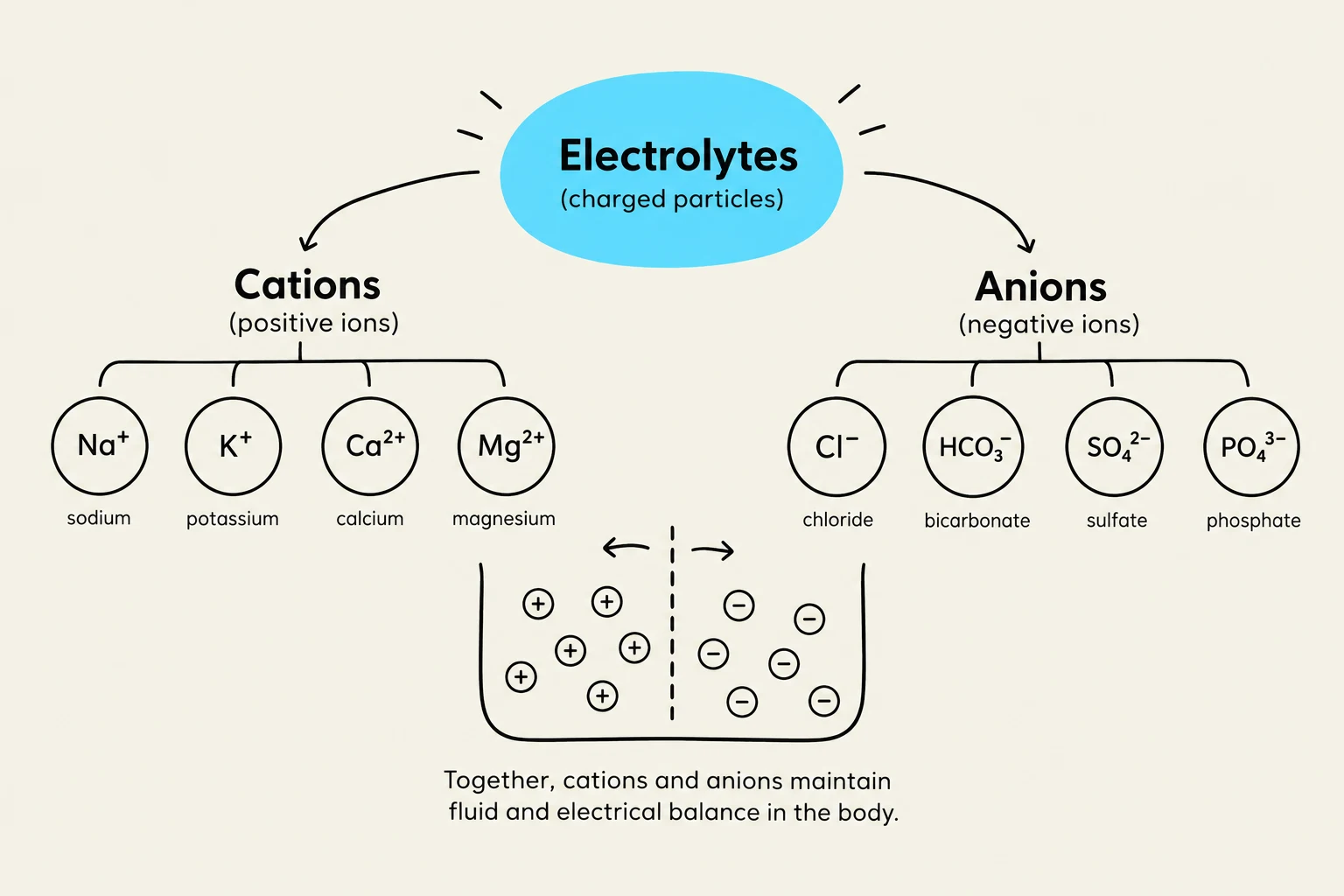
This diagram illustrates electrolytes, which are charged particles. It categorizes them into cations (positive ions) like sodium, potassium, calcium, and magnesium, and anions (negative ions) such as chloride, bicarbonate, sulfate, and phosphate. The image also shows that cations and anions together maintain fluid and electrical balance in the body.
Fluid Movement and Osmolality
Fluid does not move randomly; it moves according to specific physiological mechanisms:
Diffusion: The passive movement of particles from an area of high concentration to low concentration.
Osmosis: The movement of water across a semi-permeable membrane.
Filtration: The process by which water and solutes move across a cell membrane.
Active Transport: Requires energy in the form of ATP to move electrolytes from a low to a high concentration (e.g., the Na+/K+ pump).
A critical concept in fluid movement is osmolality, which measures the concentration of solute particles per kilogram of water. This determines the tonicity of a fluid, which dictates how water moves in relation to cells:
Tonicity
Description
Effect on Cells
Isotonic
Same concentration as blood
No net water movement; stays in the intravascular space.
Hypotonic
More dilute than blood
Water moves INTO the cells.
Hypertonic
More concentrated than blood
Water moves OUT of the cells.
Exam Tip: The "Tonicity Trick"
Isotonic = Stay: Fluid stays in the bloodstream.
Hypotonic = In: Water moves into the cells.
Hypertonic = Out: Water moves out of the cells.
Fluid Imbalances: Hypervolemia and Hypovolemia
When fluid intake and output are not synchronized, imbalances occur.
Hypervolemia (Excessive Fluid Volume)
This occurs when there is increased fluid retained in the intravascular compartment. Common causes include heart failure, kidney failure, or cirrhosis.
Also known as dehydration, this occurs when fluid loss exceeds intake. Common causes include vomiting, diarrhea, excessive sweating, or fever.
Early Signs: Tachycardia, increased capillary refill (>2 seconds), dry mucous membranes, and dizziness.
Late Signs: Hypotension, bradycardia, decreased consciousness, and anuria (lack of urine).
This illustration compares hypervolemia (too much fluid in the body) with hypovolemia (too little fluid in the body), detailing associated symptoms such as edema and JVD for hypervolemia, and hypotension, tachycardia, and dry mucous membranes for hypovolemia.
Acid-Base Balance and ABG Interpretation
The body must maintain a blood pH between 7.35 and 7.45. If the pH falls below 7.35, the patient is in acidosis; if it rises above 7.45, they are in alkalosis.
The two primary systems regulating pH are the Lungs (Respiratory) and the Kidneys (Metabolic):
Respiratory System: Controls CO2 levels. A fast response (minutes) via exhalation of carbonic acid.
Renal System: Controls bicarbonate (HCO3−) and hydrogen ions (H+). A slow response (hours to days).
Interpreting Arterial Blood Gases (ABGs)
To determine the cause of an imbalance, clinicians use a 4-step approach:
Check pH: Is it Acidosis (<7.35) or Alkalosis (>7.45)?
Check PaCO2 (Respiratory): Normal is 35–45 mmHg.
Check HCO3− (Metabolic): Normal is 22–26 mEq/L.
Determine Compensation: Is the other system trying to fix the pH?
Condition
pH
PaCO2
HCO3−
Respiratory Acidosis
↓
↑
Normal
Respiratory Alkalosis
↑
↓
Normal
Metabolic Acidosis
↓
Normal
↓
Metabolic Alkalosis
↑
Normal
↑
Memory Trick for Compensation:
Uncompensated: Only one system (pH, CO2, or HCO3−) is abnormal.
Partially Compensated: Both CO2 and HCO3− are abnormal, but pH is still not normal.
Fully Compensated: pH has returned to the normal range (7.35–7.45).
Clinical Application: IV Therapy Calculations
In clinical practice, managing these balances often requires Intravenous (IV) therapy. Nurses must be proficient in calculating flow rates for both electronic pumps and gravity lines.
1. IV Pump (mL/hr)
The pump is the most accurate method for delivering precise doses.
Formula:Flow Rate (mL/hr)=Total Time (hr)Total Volume (mL)
Example: An order for 1000 mL of Normal Saline to infuse over 8 hours.
mL/hr=8 hr1000 mL=125 mL/hr
2. Gravity Infusion (gtt/min)
When using a manual roller clamp, you must calculate the "drip rate" based on the tubing's drop factor.
Formula:Flow Rate (gtt/min)=Total Time (minutes)Total Volume (mL)×Drop Factor (gtt/mL)
Example: Infuse 1000 mL over 8 hours using tubing with a drop factor of 15 gtt/mL.
First, convert hours to minutes: 8×60=480 minutes.
gtt/min=4801000×15=48015000=31.25≈31 gtt/min
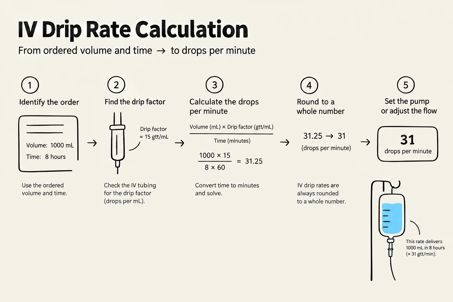
This diagram illustrates the calculation of IV drip rates, converting ordered volume and time into drops per minute. It outlines five steps: identifying the order, finding the drip factor, calculating drops per minute using a formula, rounding to a whole number, and setting the pump or adjusting the flow.
Summary Checklist for Nurses:
Monitor: Daily weights, I&O, lung sounds, and electrolytes.
Assess IV Sites: Watch for redness (phlebitis), coolness/swelling (infiltration), or heat (infection).
Safety: Always "Scrub the Hub" and verify provider orders before initiating any infusion.
Keep reading for free.
Create an account to finish this reading and make it part of your study routine.