Understanding Hemolysis Patterns on Blood Agar for Bacterial Identification
This guide explains the three types of hemolysis (Alpha, Beta, and Gamma) observed on blood agar plates. It details how these patterns, indicating the breakdown of red blood cells by bacterial hemolysins, are used in microbiology to differentiate bacteria and assess their pathogenic potential. The content covers the vi
In the microbiology laboratory, the ability to distinguish between different species of bacteria often relies on observing how they interact with their environment. One of the most fundamental and visually striking methods used for this purpose is the observation of hemolysis on blood agar. By observing how bacteria break down red blood cells, microbiologists can gain immediate clues regarding the organism's identity and its potential to cause disease in a human host.
What is Blood Agar?
Blood agar is a type of enriched, differential medium. It consists of a basal medium (such as Tryptic Soy Agar) supplemented with a specific concentration of mammalian blood, most commonly sheep blood.
Because it contains red blood cells (RBCs), it serves two primary functions:
Enriched Medium: It provides extra nutrients (like hemoglobin and vitamins) that allow fastidious organisms—those with complex nutritional requirements—to grow.
Differential Medium: It allows us to differentiate bacteria based on their ability to produce hemolysins. Hemolysins are extracellular enzymes (toxins) produced by bacteria that damage or destroy the membranes of red blood cells.
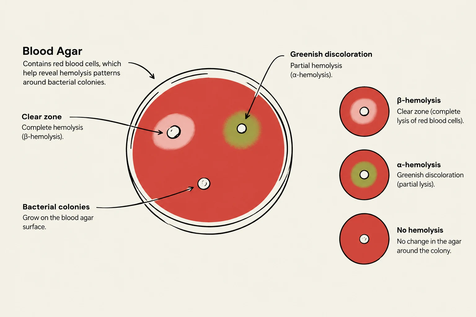
This illustration shows blood agar, a culture medium used in microbiology to grow bacteria. It demonstrates different types of hemolysis (red blood cell lysis) that can occur around bacterial colonies, including beta-hemolysis (complete lysis, indicated by a clear zone), alpha-hemolysis (partial lysis, indicated by greenish discoloration), and no hemolysis (no change in the agar).
The Three Patterns of Hemolysis
When bacteria are streaked onto a blood agar plate, the resulting growth pattern is categorized into three distinct types: Alpha, Beta, and Gamma hemolysis. Understanding these patterns is a cornerstone of clinical diagnostics.
1. Alpha (α) Hemolysis: Partial Destruction
Alpha hemolysis is characterized by a partial breakdown of red blood cells. Instead of a clear zone, you will observe a greenish or brownish discoloration of the agar immediately surrounding the bacterial colonies.
This color change is not actually due to the destruction of the cells themselves, but rather the oxidation of hemoglobin into methemoglobin. Because the red blood cells are not completely lysed, the agar remains opaque, just with a different hue.
Exam Tip: If you see a "bruised" or greenish look around a colony, think Alpha hemolysis. It indicates partial damage, not total destruction.
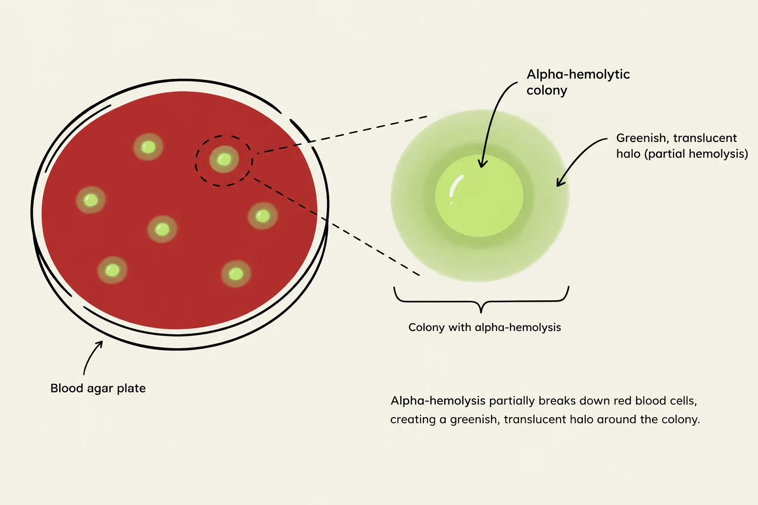
Illustration showing alpha-hemolysis on a blood agar plate, where bacterial colonies partially break down red blood cells, creating a greenish halo.
2. Beta (β) Hemolysis: Complete Destruction
Beta hemolysis is the most dramatic pattern. It is characterized by the complete lysis (rupture) of red blood cells and the total destruction of hemoglobin in the area surrounding the colony.
On a plate, this appears as a clear, transparent zone around the bacterial growth. If you hold the plate up to the light, you can see right through the agar where the bacteria have grown. This pattern is often associated with highly virulent pathogens, such as Streptococcus pyogenes, which produces potent toxins to invade host tissues.
3. Gamma (γ) Hemolysis: No Hemolysis
Gamma hemolysis (sometimes referred to as non-hemolytic) occurs when the bacteria grow on the surface of the agar but do not produce any hemolysins.
In this case, there is no change in the color or transparency of the medium. The agar remains its original opaque red color right up to the edge of the bacterial colony. While "gamma" implies a lack of hemolytic activity, it is important to remember that these organisms may still be pathogenic through other mechanisms, such as toxin production that does not target RBCs.
This diagram illustrates the different types of hemolysis (Alpha, Beta, and Gamma) observed on blood agar plates, which is a method used for bacterial identification based on their ability to lyse red blood cells.
Clinical Significance and Pathogenic Potential
The pattern of hemolysis is more than just a visual exercise; it is a vital tool for assessing the "pathogenic potential" of an unknown isolate.
Hemolysis Type
Visual Appearance
Mechanism
Pathogenic Implication
Alpha (α)
Greenish/Brownish discoloration
Partial oxidation of hemoglobin
Often less virulent, but can be significant (e.g., certain Streptococcus pneumoniae)
Beta (β)
Clear, transparent zone
Complete lysis of RBCs
Often indicates high virulence and invasive potential
Gamma (γ)
No change in agar
No hemolysin production
Variable; may be commensal or pathogenic via other routes
In a clinical setting, if a patient presents with a severe infection and the lab identifies a β-hemolytic organism, the clinician can immediately suspect a highly aggressive pathogen. This rapid differentiation allows for faster decision-making in prescribing antibiotics and implementing infection control measures.
Summary for Students
To master this topic, remember the "Total vs. Partial" rule:
Beta = Total destruction (Clear zone).
Alpha = Partial destruction (Green zone).
Gamma = None (No change).
When performing these tests in the lab, always ensure you are observing the plate against a light source to accurately distinguish between the transparency of Beta hemolysis and the slight discoloration of Alpha hemolysis.
Keep reading for free.
Create an account to finish this reading and make it part of your study routine.