Understanding Hemagglutination: Principles and Applications
This reading explains the principles of hemagglutination, a technique using red blood cells to detect antigens and antibodies. It covers its mechanisms, differentiation from hemolysis, use of conjugated antigens, titration for quantification, and clinical applications like blood typing and viral detection.
In the field of immunology and diagnostic medicine, detecting the presence of specific antigens is a fundamental task. One of the most elegant and visually intuitive methods used to achieve this is hemagglutination. This technique leverages the unique physical properties of red blood cells (RBCs) to act as visual indicators for the presence of specific antigens or antibodies. By observing whether red blood cells clump together or settle individually, scientists can determine the presence, concentration, and affinity of target molecules.
The Fundamental Principle of Hemagglutination
At its core, hemagglutination is a phenomenon where red blood cells aggregate into a lattice-like structure. This occurs when an agent—typically an antibody or a lectin—acts as a bridge between the antigens located on the surfaces of multiple red blood cells.
To understand this, we must look at the surface of the erythrocyte (red blood cell). The membrane of an RBC is studded with various glycoproteins and glycolipids that serve as antigens. When a multivalent molecule (a molecule with multiple binding sites) is introduced into a solution containing these cells, it can bind to an antigen on one cell and simultaneously bind to an antigen on a neighboring cell.
Diagram illustrating the difference between dispersed red blood cells and agglutinated red blood cells forming a lattice structure.
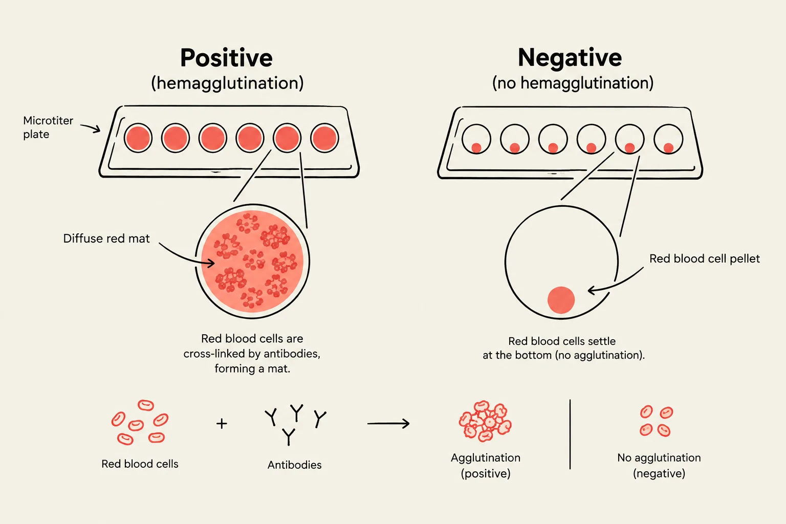
If the antigen is present and the bridging agent is functional, the cells will form a large, interconnected network. This network is heavy enough to stay suspended or create a visible "mat" at the bottom of a testing well, rather than settling into a tight, smooth pellet.
Mechanisms of Detection: Agglutination vs. Hemolysis
It is crucial for students to distinguish between hemagglutination and hemolysis, as these terms are often confused in exam settings.
Hemagglutination: This is the physical clumping of cells. The integrity of the red blood cell membrane remains intact. The cells are simply "stuck" together in a group.
Hemolysis: This refers to the rupture of the red blood cell membrane, causing the release of hemoglobin into the surrounding fluid. This results in a clear, red-colored liquid rather than a clumped suspension.
Exam Tip: If a question asks about the "clumping" of cells, think agglutination. If it asks about the "destruction" or "bursting" of cells, think hemolysis.
Using Conjugated Antigens for Detection
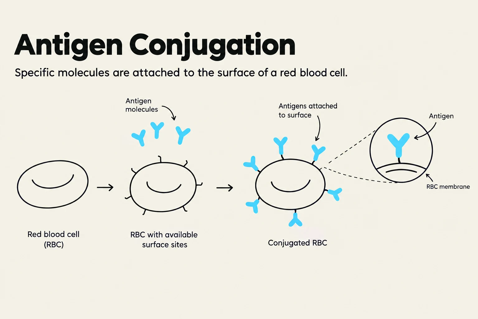
While natural hemagglutination often involves antibodies reacting to natural blood group antigens, a highly controlled version of this technique involves using conjugated antigens. In this variation, specific antigens are chemically attached (conjugated) to the surface of red blood cells to turn the cells into "reporter" particles.
In this setup, the red blood cell is no longer just a biological sample; it is a tool. By coating an RBC with a specific antigen, you can use it to detect the presence of a specific antibody in a patient's serum. If the patient's serum contains antibodies that recognize that conjugated antigen, the cells will agglutinate.
This diagram illustrates the process of antigen conjugation, where specific molecules attach to the surface of a red blood cell (RBC).
The Mathematical Logic of Titration
To quantify how much antibody is present in a sample, scientists use a process called titration. This involves performing a serial dilution of the sample and observing the highest dilution at which agglutination still occurs.
If we define the concentration of the antibody as C and the dilution factor as D, the effective concentration in a specific well can be expressed as:
Cwell=DCinitial
By finding the point where the "clumping" disappears, we can determine the titer, which is a standard measure of the concentration of an antigen or antibody in a biological sample.
Clinical and Laboratory Applications
Hemagglutination is not merely a theoretical concept; it is a workhorse in diagnostic laboratories.
Blood Typing: The most common application is determining ABO and Rh blood groups. By exposing a patient's blood to known antibodies (anti-A, anti-B, etc.), clinicians can observe agglutination to identify the patient's blood type.
Viral Detection: Many viruses, such as the influenza virus, possess hemagglutinin proteins on their envelopes. These proteins naturally cause red blood cells to clump. Measuring this clumping can help quantify the amount of virus present in a sample.
Autoimmune Screening: Detecting if a patient's immune system is producing antibodies against their own red blood cells (autoantibodies).
Diagram illustrating hemagglutination (positive) versus no hemagglutination (negative) in a microtiter plate, showing the interaction of red blood cells and antibodies.
Summary Table for Quick Review
Feature
Hemagglutination
Hemolysis
Cell Membrane
Intact
Ruptured
Visual Result
Clumping/Lattice formation
Clear red liquid (hemoglobin release)
Primary Cause
Bridging by antibodies/lectins
Complement activation or toxins
Diagnostic Use
Detecting antigens/antibodies
Detecting cell-destructive agents
By mastering the visual cues and the underlying molecular mechanics of red blood cell clumping, students can effectively interpret immunological assays and understand the vital role of antigen-antibody interactions in clinical diagnostics.
Keep reading for free.
Create an account to finish this reading and make it part of your study routine.