Understanding Graded and Voltage-Gated Potentials in Excitable Cells
This content explains the fundamental differences between graded potentials and action potentials in excitable cells, detailing the roles of chemically-gated and voltage-gated ion channels in cellular communication and signal transmission.
In the study of neurobiology and muscle physiology, understanding how cells communicate requires a deep dive into electrical signals. These signals, known as membrane potentials, are not uniform; they vary in how they are triggered, how far they travel, and how they change the electrical state of the cell. By distinguishing between graded potentials and voltage-gated potentials, we can understand how a single chemical signal at a synapse can eventually trigger a massive muscle contraction or a nerve impulse.
Graded Potentials: Localized Signals
Graded potentials are the "first responders" of the cellular electrical system. They are typically produced by the opening of chemically gated ion channels. When a specific chemical (a ligand) binds to a receptor on the cell membrane, these channels open, allowing ions to flow into or out of the cell.
Unlike the powerful action potentials that follow, graded potentials possess two defining characteristics:
Local: These potentials are restricted to the specific area of the membrane where the stimulus occurred, such as the dendrites or the soma of a neuron, or the motor end plate of a muscle cell.
Decremental: As the electrical change moves away from the initial site of the stimulus, its magnitude decreases. This is because the ions diffuse through the cytosol and spread out, losing their concentrated effect as they travel.
Because they are decremental, a single graded potential might not be strong enough to trigger a response. However, if many chemical gates open simultaneously, or if a gate stays open for a long time, the cumulative effect can reach a critical level known as threshold.
Exam Tip: If a question asks about the difference between a graded potential and an action potential, remember that graded potentials are decremental (they fade away) and local, whereas action potentials are non-decremental and propagate (travel long distances without losing strength).
This diagram illustrates the concept of graded potentials in neurons, showing how different stimulus strengths lead to varying degrees of membrane depolarization. It depicts a synapse where a stimulus triggers ion flow across the cell membrane, resulting in changes in membrane potential, with larger stimuli causing larger changes.
Voltage-Gated Ion Channels
While graded potentials rely on chemicals to open their gates, voltage-gated ion channels are much more selective. These membrane proteins respond specifically to changes in the membrane potential—the difference in electrical charge between the inside and the outside of the cell.
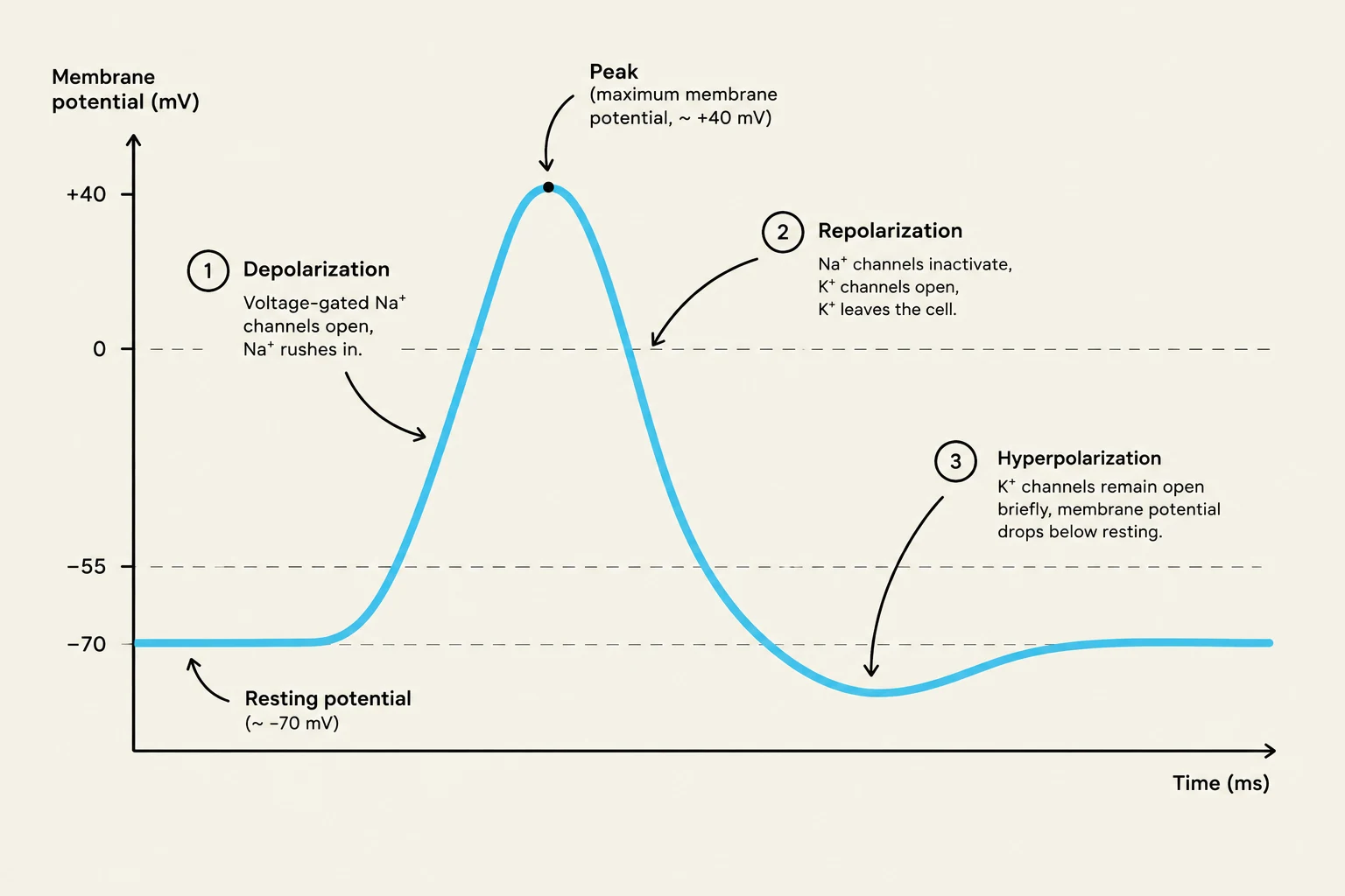
The behavior of these channels is dictated by the cell's current electrical state. For example, at the resting membrane potential, a neuron typically maintains a charge of approximately −70 mV, while a skeletal muscle cell sits at approximately −90 mV. Under these resting conditions, the voltage-gated channels responsible for the action potential remain closed.
The Role of Voltage-Gated Potassium Channels
One of the most critical players in resetting the cell's electrical state is the voltage-gated potassium (K+) channel. These channels follow a specific cycle:
At Rest: The channels are closed.
At Maximum Potential: When the cell reaches its peak electrical charge (the maximum membrane potential, often around +30 mV), these channels snap open.
Potassium Efflux: Once open, potassium ions rush out of the cell, a process called efflux. Because positive ions are leaving, the inside of the cell becomes more negative again.
Repolarization: This drop in membrane potential back toward a negative value is known as repolarization.
Slow Closure and Hyperpolarization: These channels are "slow to close"—they don't slam shut like a door, but rather "soft close". Because they remain slightly open even as the cell approaches its resting state, a small amount of potassium continues to leak out, causing the potential to dip slightly below the resting level. This temporary dip is called hyperpolarization.
A graph illustrating the changes in membrane potential during an action potential, showing depolarization, repolarization, and hyperpolarization phases.
From Threshold to Action Potential
The transition from a local graded potential to a full-scale action potential occurs at the axon hillock (in neurons) or the motor end plate (in muscle cells). This area is densely packed with voltage-gated channels.
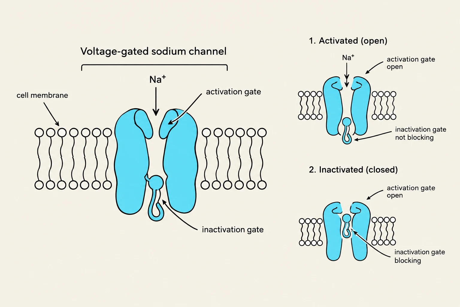
When the sum of all incoming graded potentials pushes the membrane potential to the threshold, it triggers the voltage-gated sodium (Na+) channels. These channels are unique because they have two gates: an activation gate and an inactivation gate.
Depolarization: At threshold, the activation gate opens, allowing a massive sodium influx (sodium rushing into the cell). This causes the membrane potential to shoot upward rapidly toward the maximum potential.
The Peak: Once the maximum potential is reached, the sodium channel's inactivation gate closes, stopping the flow of sodium.
The Reset: As the cell repolarizes via potassium efflux, the sodium channel eventually "resets"—the activation gate closes and the inactivation gate opens, making the channel ready for the next signal.
Note on Refractory Periods: During the time when sodium channels are inactivated and cannot be reopened, the cell is in an absolute refractory period. This ensures that the action potential only travels in one direction and cannot move backward.
Diagram illustrating the structure and function of a voltage-gated sodium channel in a cell membrane, showing its activated (open) and inactivated (closed) states.
Summary Table: Comparing Potentials
To help consolidate these concepts, refer to the following comparison of the two primary electrical events in excitable cells:
Feature
Graded Potential
Voltage-Gated Potential (Action Potential)
Trigger
Chemical/Ligand binding
Change in membrane voltage (Threshold)
Distance
Local/Short-range
Long-range/Propagated
Magnitude
Decremental (fades with distance)
Non-decremental (constant strength)
Primary Channels
Chemically-gated channels
Voltage-gated Na+ and $K^+$ channels
By understanding these mechanisms, we can see how the body converts a chemical message into a precise, traveling electrical impulse that can trigger everything from a thought to a muscle movement.
Keep reading for free.
Create an account to finish this reading and make it part of your study routine.