Maintaining a stable internal environment is a fundamental requirement for cellular function. This state, known as homeostasis, relies on the precise balance of fluids and electrolytes. The human body organizes its water into distinct compartments, primarily the Intracellular Fluid (ICF)—the fluid inside the cells—and the Extracellular Fluid (ECF)—the fluid outside the cells. Understanding how water moves between these spaces, as well as between the blood vessels (plasma) and the surrounding tissues (interstitial fluid), is essential for recognizing clinical imbalances like edema, dehydration, and shock.
The Fluid Compartments and Tonicity
The body's total body water (TBW) is distributed unevenly between the ICF and ECF. These compartments are separated by semi-permeable membranes, such as cell membranes and capillary walls, which allow certain substances to pass while restricting others.
The chemical composition of these compartments is highly specific:
Intracellular Fluid (ICF): Characterized by high concentrations of Potassium ($K^+$), Magnesium (Mg2+), Phosphate (PO43−), and proteins.
Extracellular Fluid (ECF): Characterized by high concentrations of Sodium (Na+), Chloride ($Cl^-$), and Bicarbonate (HCO3−).
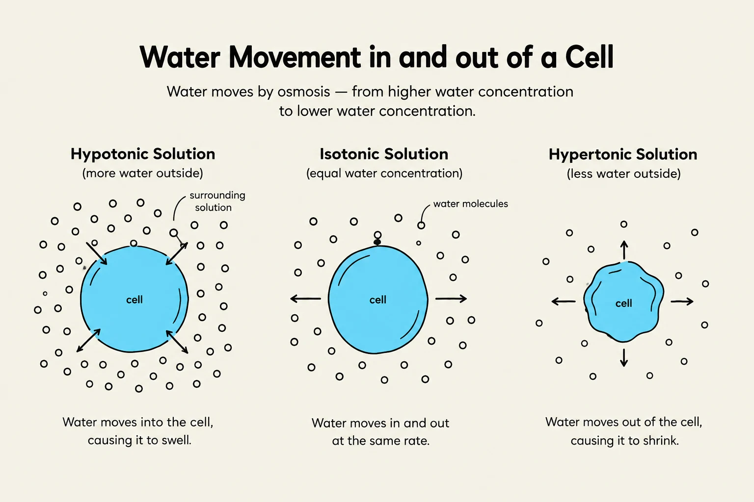
Because these compartments have different concentrations of solutes, water moves between them via osmosis—the movement of water from an area of low solute concentration to an area of high solute concentration. The direction of this movement is determined by the tonicity of the solution:
Isotonic: The solute concentration is equal inside and outside the cell. There is no net movement of water.
Hypotonic: The solution has a lower solute concentration than the cell. Water moves into the cell, causing it to swell.
Hypertonic: The solution has a higher solute concentration than the cell. Water moves out of the cell, causing it to shrink.
This diagram illustrates the movement of water into and out of a cell through osmosis, depending on the concentration of the surrounding solution. It shows hypotonic, isotonic, and hypertonic solutions and their effects on cell volume.
Exam Tip: When thinking about tonicity, remember that "water follows salt." If a solution is hypertonic (high salt), water will leave the cell to try and dilute that salt.
Capillary Exchange: Starling Forces
While osmosis governs movement between cells and the ECF, a different set of physical forces governs the exchange of fluid between the blood plasma and the interstitial space (the fluid surrounding the cells). This process is known as Net Filtration, and it is determined by the balance of Starling forces.
The formula for net filtration is:
Net Filtration=(Forces favoring filtration)−(Forces opposing filtration)
Forces Favoring Filtration (Pushing water out of the capillary)
Capillary Hydrostatic Pressure: This is essentially the blood pressure within the capillary. It acts as a "pushing" force that drives water out of the vessel and into the interstitial space.
Interstitial Oncotic Pressure: This is the osmotic pressure exerted by solutes (like proteins) in the interstitial space that "pulls" water out of the capillary.
Forces Favoring Reabsorption (Pulling water back into the capillary)
Capillary Oncotic Pressure: This is a powerful "pulling" force exerted by proteins, most notably albumin, located within the plasma. Because proteins are too large to easily cross the capillary wall, they create an osmotic gradient that keeps water inside the blood vessel.
Interstitial Hydrostatic Pressure: The pressure exerted by the fluid in the interstitial space that helps facilitate the inward movement of water toward the capillary.
Diagram illustrating capillary hydrostatic pressure and capillary oncotic pressure, showing fluid movement into and out of the capillary.
Clinical Imbalances: Edema and Volume Shifts
When the delicate balance of Starling forces or tonicity is disrupted, fluid accumulates in places it shouldn't be, or disappears from where it is needed.
Edema Formation
Edema is the accumulation of fluid in the interstitial spaces. This occurs through several primary mechanisms:
Increased Capillary Hydrostatic Pressure: Often caused by heart failure or high blood pressure, which "pushes" too much fluid out of the vessels.
Decreased Plasma Oncotic Pressure: If a patient has low levels of albumin (due to liver failure or kidney disease/nephrotic syndrome), there isn't enough "pull" to keep water in the vessels.
Increased Capillary Permeability: During inflammation or burns, capillary walls become "leaky," allowing fluid and proteins to escape into the tissues.
Lymphatic Obstruction: If lymph channels are blocked by tumors, infection, or surgery, the fluid that normally drains from the tissues cannot return to the circulation, leading to lymphedema.
Edema can be localized or generalized. Severe, widespread edema throughout the entire body is referred to as anasarca. Clinically, providers often check for pitting edema, where pressing a finger into the swollen area leaves a persistent indentation.
Fluid Volume Deficit (FVD) and Excess
Fluid imbalances are categorized by how they affect the total volume and the concentration of electrolytes:
Extracellular Fluid Volume Deficit (FVD): Occurs when there is insufficient fluid to meet metabolic needs.
Isotonic loss: Loss of both water and sodium in equal proportions.
Hypotonic loss: Loss of more sodium than water.
Hypertonic loss: Loss of more water than sodium.
Dehydration: Specifically refers to the loss of water without a corresponding loss of sodium.
Severe FVD can lead to hypovolemic shock, a critical state where inadequate blood volume results in poor tissue perfusion.
Extracellular Fluid Volume Excess: Results from increased intake, fluid retention, or shifts from the interstitial space into the intravascular space.
Regulation and Lifespan Considerations
The body uses complex hormonal systems to maintain this balance. The Renin-Angiotensin-Aldosterone System (RAAS) and Antidiuretic Hormone (ADH) are central to renal regulation. Aldosterone promotes sodium and water reabsorption, while ADH increases water reabsorption in the kidneys to prevent water loss. Additionally, natriuretic peptides act as a counterbalance by telling the kidneys to excrete sodium and water to lower blood volume.
Age significantly impacts fluid stability:
Infants: Have a much higher percentage of total body water and a larger body surface area relative to their mass, making them highly susceptible to rapid dehydration.
Older Adults: Often have decreased thirst perception, reduced renal function (lower GFR), and decreased muscle mass, increasing their risk for both dehydration and acute kidney injury (AKI).
Failure to manage these fluid and electrolyte shifts can lead to catastrophic outcomes, including Acute Kidney Injury (AKI), Chronic Kidney Disease (CKD), and Multiple Organ Dysfunction Syndrome (MODS).
Keep reading for free.
Create an account to finish this reading and make it part of your study routine.