Understanding Fluid and Electrolyte Balance in Nursing
A comprehensive guide for nurses on maintaining fluid and electrolyte balance, covering fluid compartments, regulatory systems, imbalances, and nursing assessment with IV therapy.
Maintaining homeostasis—a stable internal balance—is one of the most critical functions of the human body. For nurses, understanding fluid and electrolyte balance is not just about memorizing numbers; it is about recognizing early risk, connecting assessment cues to specific imbalances, and implementing safe, patient-centered interventions.
Fluid Compartments and Movement
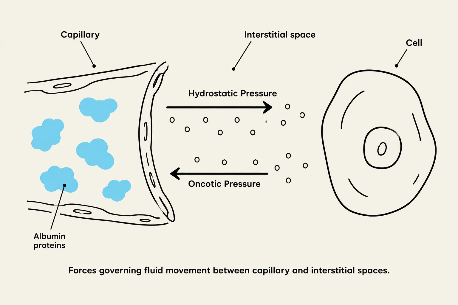
Diagram illustrating hydrostatic and oncotic pressure forces governing fluid movement between a capillary and interstitial space, with albumin proteins shown within the capillary.
Fluid in the body is distributed between two primary compartments: the intracellular fluid (ICF), which is the fluid inside the cells and represents the largest compartment, and the extracellular fluid (ECF). The ECF is further divided into interstitial fluid (between cells), intravascular fluid (plasma inside blood vessels), and transcellular fluid (specialized spaces like cerebrospinal or pleural fluid).
Movement between these compartments is driven by several physical processes:
Osmosis: Water moves toward the side with a higher effective solute concentration across a membrane.
Diffusion: Particles move from an area of high concentration to low concentration.
Filtration: Fluid and small solutes move due to pressure gradients.
Active Transport: Substances move against a concentration gradient, requiring energy (ATP).
In the capillary beds, two specific pressures dictate fluid movement. Hydrostatic pressure pushes fluid out of the capillaries, while oncotic pressure (driven by plasma proteins like albumin) acts as a "pulling" force to retain fluid in the bloodstream.
Clinical Connection: If a patient has low albumin (common in liver disease), oncotic pressure decreases, which can allow fluid to leak into the tissues, causing edema even if the patient's circulating blood volume is actually low.
Regulatory Systems
The body uses complex feedback loops to maintain balance. When circulating volume or blood pressure drops, the renin-angiotensin-aldosterone system (RAAS) is activated. This leads to vasoconstriction and the release of aldosterone, which promotes sodium and water reabsorption while excreting potassium.
Simultaneously, antidiuretic hormone (ADH) is released to increase water reabsorption in the kidneys, reducing urine output to save water. Conversely, when the atria of the heart are stretched due to high volume, atrial natriuretic peptide (ANP) is released to promote the excretion of sodium and water.
Fluid Imbalances: Deficit vs. Excess
Nurses must distinguish between volume (the amount of fluid) and concentration (the amount of solute relative to water).
Hypovolemia (Deficit)
Caused by vomiting, diarrhea, hemorrhage, or excessive diuretics, hypovolemia results in reduced circulating volume.
Labs: May show increased BUN and hematocrit due to concentration.
Hypervolemia (Excess)
Often caused by heart, kidney, or liver failure, this involves sodium and water retention.
Assessment Cues: Hypertension, bounding pulses, jugular vein distention (JVD), edema, and crackles in the lungs.
Priority: New-onset dyspnea or crackles during fluid replacement is a medical emergency indicating circulatory overload.
Third Spacing: This occurs when fluid is trapped in a space where it is not physiologically useful (such as the peritoneal cavity in ascites), which can coexist with signs of both deficit and excess.
Electrolyte Imbalances
Electrolytes are charged particles that are essential for nerve conduction, muscle contraction, and cardiac rhythm.
Sodium (Na+) [136–145 mEq/L]
Sodium primarily affects neurological status.
Hyponatremia (<136): Can cause brain swelling, headache, confusion, seizures, and decreased consciousness.
Hypernatremia (>145): Causes cellular dehydration, thirst, and irritability.
Potassium (K+) [3.5–5.0 mEq/L]
Potassium is the most critical electrolyte for cardiac rhythm.
Hypokalemia (<3.5): Causes muscle weakness, cramps, and increased risk of digoxin toxicity.
Hyperkalemia (>5.0): Can lead to life-threatening dysrhythmias or cardiac arrest.
CRITICAL SAFETY ALERT: NEVER IV PUSH OR BOLUS POTASSIUM. It must be diluted and administered via a controlled infusion pump.
Calcium (Ca2+) [9–10.5 mg/dL] and Magnesium (Mg2+) [1.3–2.1 mEq/L]
These two electrolytes often work together to regulate neuromuscular excitability.
Low Calcium/Magnesium: Leads to increased excitability, tremors, and tetany. Look for Chvostek’s sign (facial twitch) and Trousseau’s sign (hand spasm).
High Magnesium: Depresses the central nervous system, leading to reduced deep tendon reflexes, hypotension, and respiratory depression.
Other Electrolytes
Chloride (Cl−) [98–106 mEq/L]: Often follows sodium and fluid changes.
Phosphorus (PO43−) [3–4.5 mg/dL]: Influenced by nutrition and renal function; often moves inversely with calcium.
Nursing Assessment and IV Therapy
Accurate Measurement
The most reliable way to monitor fluid status is through daily weights and Intake and Output (I&O).
Weight: A change of 1 kg≈1 L of fluid. Always weigh at the same time, on the same scale, with similar clothing.
I&O: Include all liquids, including ice chips (count as half their volume) and enteral feedings.
IV Fluid Types
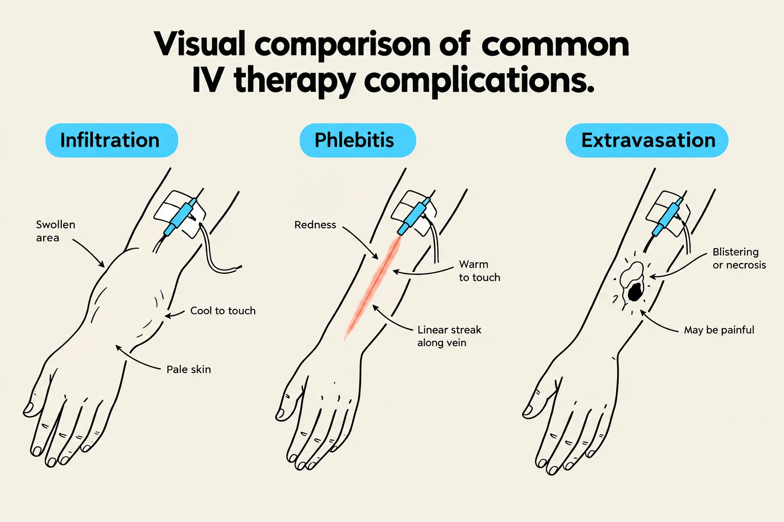
This diagram illustrates and compares three common complications of IV therapy: infiltration, phlebitis, and extravasation, detailing their visual and tactile symptoms.
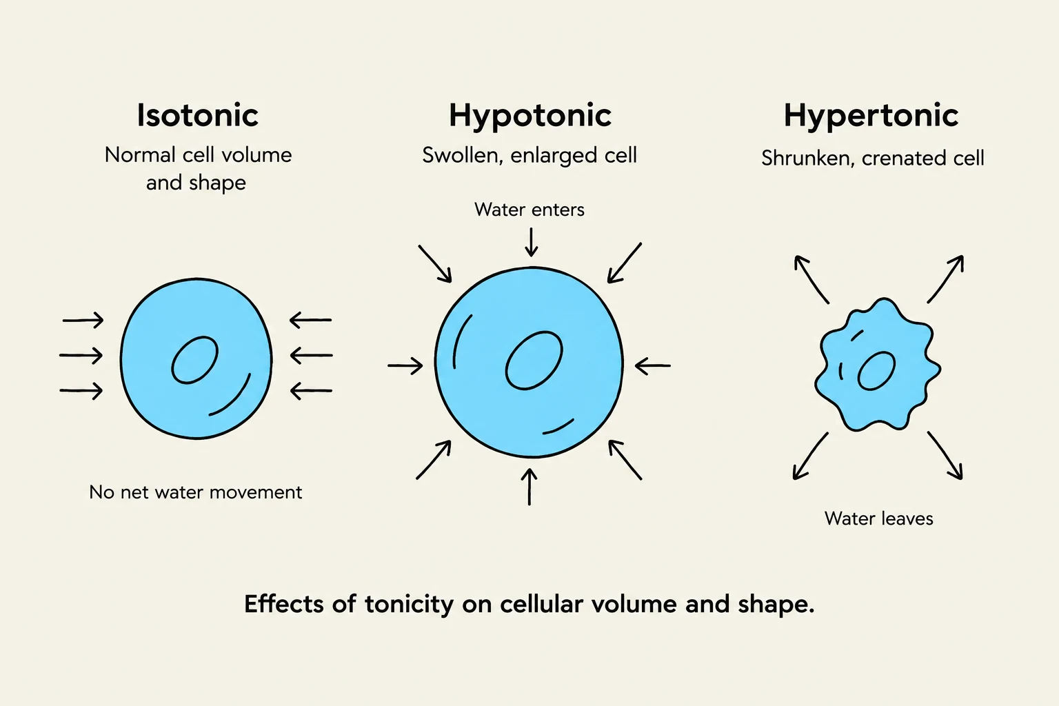
Nurses must understand the tonicity of ordered fluids to prevent cellular damage:
Isotonic (e.g., 0.9% NS, Lactated Ringer's): Expands the extracellular/intravascular volume. Monitor for circulatory overload.
Hypotonic (e.g., 0.45% NS): Causes water to move into the cells, making them swell. Monitor for neurological changes.
Hypertonic (e.g., 3% Saline): Draws water out of the cells into the ECF, shrinking the cells. Requires close monitoring for vein irritation and overload.
Colloids (e.g., Albumin): Large molecules that stay in the intravascular space to support oncotic pressure.
IV Complications
Infiltration: Cool, swollen, and pale site; non-vesicant fluid has leaked into tissue.
Extravasation: Leakage of a vesicant (irritating) medication; carries high risk for tissue injury.
Phlebitis: Redness, warmth, and pain along the vein.
Nursing Tip: If you suspect an IV complication, stop the infusion immediately. For infiltration, assess the site and follow facility policy for catheter removal.
This diagram illustrates the effects of tonicity on cellular volume and shape, showing how cells respond in isotonic, hypotonic, and hypertonic environments.
Summary of Laboratory Trends
When evaluating a patient, never rely on a single lab value. Look for trends in:
Serum Osmolality (285–295 mOsm/kg): Measures the concentration of particles in the plasma.