Understanding Epilepsy: Electrical Neurotransmission and Seizure Mechanisms
Explores the neurological disorder of epilepsy, detailing the ionic basis of action potentials, mechanisms of control like inactivation and refractory periods, and the balance of neurotransmitters (glutamate vs. GABA) that lead to seizures. It also covers seizure classification.
Epilepsy is a neurological disorder characterized by recurrent, unprovoked seizures, which are transient alterations in brain function caused by abnormal electrical activity in the cerebral cortex. While a single episode of abnormal electrical discharge is termed a "seizure," epilepsy refers to the chronic predisposition to these events. At its core, epilepsy represents a breakdown in the delicate balance of excitatory and inhibitory neurotransmission, leading to the hypersynchronous and repetitive firing of neuronal populations.
The Ionic Basis of the Action Potential
To understand how seizures occur, we must first understand how a normal neuron communicates. Nerve signaling relies on the movement of ions across the cell membrane through specialized channels. When a neuron is stimulated, it undergoes membrane depolarization, a process where the internal charge of the cell becomes more positive.
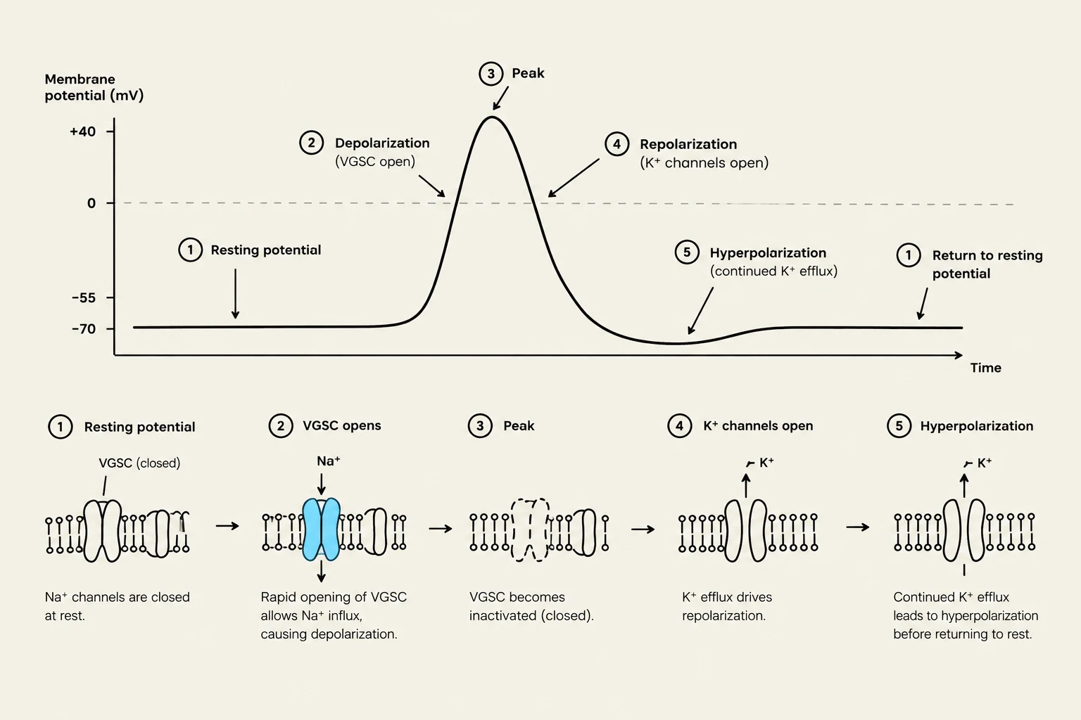
This depolarization is driven primarily by voltage-gated Na+ channels (VGSC). When the membrane potential reaches a certain threshold, these channels open rapidly, allowing an influx of sodium ions (Na+) into the cell, which propagates the action potential along the membrane. To prevent the cell from remaining in a permanent state of excitation, voltage-gated K+ channels (VGPC) open shortly after the sodium channels, allowing potassium ions ($K^+$) to exit the cell, which re-polarizes or hyper-polarizes the membrane.
This diagram illustrates the changes in membrane potential during an action potential, detailing the roles of voltage-gated sodium (Na+) and potassium (K+) channels in depolarization, repolarization, and hyperpolarization.
Mechanisms of Control: Inactivation and the Refractory Period
The brain has built-in "safety breaks" to prevent neurons from firing uncontrollably. One of the most critical mechanisms is the conformational state transition of the VGSC. These channels cycle through three states: resting, open, and inactivated.
Inactivation: As soon as the channel opens to allow sodium in, it quickly transitions into an inactivated state. This inactivation limits the duration of the action potential and ensures that the signal moves in only one direction (unidirectional propagation).
The Refractory Period: After inactivation, the channel must transition back to the resting state before it can be opened again. This recovery takes significantly longer than the initial opening process. This delay creates a refractory period, which imposes a strict "time-limit" on how frequently a single neuron can fire.
In a healthy brain, if neurons fire at a low frequency, the VGSCs have ample time to recover. However, if a group of neurons overrides these limits and begins repetitive nerve firing, they can synchronize their activity and activate neighboring neurons, potentially triggering a seizure.
Exam Tip: Remember that VGSC inactivation is the primary mechanism that prevents repetitive firing and maintains the isolation of nerve signals. If this mechanism fails, the frequency of action potentials increases, leading to synchronization.
Neurotransmitter Imbalance: Glutamate vs. GABA
The electrical stability of the brain is maintained by the balance between excitatory and inhibitory neurotransmitters.
Excitatory Transmission:Glutamate is the primary excitatory neurotransmitter. It acts on glutamate ionotropic receptors, specifically NMDA and AMPA receptors, to promote depolarization and facilitate the firing of action potentials.
Inhibitory Transmission:GABA (gamma-aminobutyric acid) is the primary inhibitory neurotransmitter. It acts on GABA type A receptors to counteract depolarization and prevent excessive excitation.
A key physiological defense against seizures is the role of GABAergic interneurons. These specialized neurons release GABA to prevent the "synchronization" of large groups of neurons. When the balance shifts—either through excessive glutamate activity or insufficient GABAergic inhibition—the brain becomes prone to the massive, synchronous electrical discharges seen in epilepsy.
Diagram illustrating synaptic transmission where glutamate released from a presynaptic neuron binds to AMPA and NMDA receptors on a postsynaptic neuron, leading to excitation. A balance scale depicts the opposing roles of excitatory glutamate and inhibitory GABA.
Classification of Seizures
Seizures are classified based on where they begin in the brain and the clinical symptoms they produce.
Focal Onset Seizures
Focal onset seizures (formerly called partial seizures) originate in a specific group of neurons limited to one part of a single cerebral hemisphere. They are further divided by the patient's level of awareness:
Focal Aware Seizures: The patient remains conscious. Symptoms depend on which cortical area is affected (e.g., sensory changes or emotions).
Focal Impaired Awareness Seizures: The patient's consciousness or contact with reality is affected. These may involve automatisms (repetitive movements like lip-smacking or hand-rubbing) or hallucinations.
Focal seizures can also evolve into focal to bilateral tonic-clonic seizures, where the activity starts in one focus and spreads to both hemispheres via the corpus callosum or thalamic projections.
Generalized Onset Seizures
Generalized onset seizures involve both cerebral hemispheres from the very beginning. These are categorized into two main types:
Absence Seizures (formerly "Petit Mal"): These are characterized by brief "staring spells" and a loss of consciousness. On an EEG, they typically show a characteristic 3-Hz spike-and-wave activity, which is linked to abnormal activity in the thalamus involving T-type Ca2+ channels.
Tonic-Clonic Seizures (formerly "Grand Mal"): These are more dramatic and involve a sudden loss of consciousness. They typically follow two phases:
Tonic phase: Sustained muscle contractions and limb extension.
Clonic phase: Rhythmic, jerking limb movements.
Following a tonic-clonic seizure, patients often experience a period of post-ictal confusion that can last from minutes to hours.
Summary of Electrical Pathophysiology
In summary, the transition from normal brain activity to a seizure involves a breakdown in the control of ion flow. The Paroxysmal Depolarization Shift (PDS) is a key electrical hallmark, characterized by high-voltage, long-lasting depolarization currents caused by a massive influx of Na+ and Ca2+ ions. When these shifts become widespread and synchronized across the cortex, the clinical manifestations of epilepsy emerge.
Keep reading for free.
Create an account to finish this reading and make it part of your study routine.