Understanding Dentition Periods and Dental Terminology
This guide explains the three dentition periods (primary, mixed, and permanent), eruption and shedding patterns, and essential dental anatomy terminology for effective clinical communication.
Understanding the progression of teeth from infancy through adulthood is a fundamental skill in clinical dentistry. A patient's "dentition" refers to the natural teeth present in the jaws. Because humans undergo two distinct sets of teeth over a lifetime, clinicians must be able to identify which dentition period a patient is currently in to provide appropriate care, from monitoring eruption patterns to planning long-term restorative treatments.
The Three Dentition Periods
While there are only two types of teeth (primary and permanent), there are three distinct clinical periods defined by which teeth are present in the mouth.
1. Primary Dentition Period
Also known as "deciduous" dentition, this period begins with the eruption of the primary mandibular central incisors. It typically spans from approximately 6 months to 6 years of age. During this stage, only primary teeth are present. These teeth are not merely "temporary" placeholders; they are critical for supporting mastication, speech, facial development, and maintaining the space necessary for future permanent teeth.
2. Mixed Dentition Period
The mixed period is a transitional phase occurring roughly between ages 6 and 12. It begins with the eruption of the first permanent tooth—most commonly the mandibular first molar—and ends when the last primary tooth is shed. During this time, both primary and permanent teeth coexist in the mouth.
Clinical Tip: During the mixed dentition period, patients may be concerned about the different colors and sizes of their teeth. Primary teeth are generally smaller and lighter in color, while permanent teeth are larger and darker.
3. Permanent Dentition Period
This period begins after the final primary tooth has been shed, typically after age 12. It is characterized by the presence of the permanent dentition, which includes the eruption of all teeth except for those that are missing or impacted (such as third molars). The focus of care shifts toward long-term maintenance, periodontal health, and occlusion.
This illustration shows the progression of human dentition from primary (baby) teeth to mixed dentition and finally to permanent (adult) teeth, detailing the number of teeth at each stage and the overlap period.
Eruption, Shedding, and Clinical Considerations
Successful dental development relies on a predictable sequence of eruption and shedding (exfoliation).
Eruption Markers and Successional Patterns
A key concept in dental anatomy is the difference between succedaneous and nonsuccedaneous teeth:
Succedaneous teeth: Permanent anterior teeth and premolars that replace their primary predecessors.
Nonsuccedaneous teeth: Permanent molars, which erupt distal to the primary dentition and do not replace any primary teeth.
Clinicians must monitor for overretained primary teeth, which are primary teeth that persist more than a year beyond their expected exfoliation time. These can interfere with the normal eruption path of permanent teeth.
Interceptive Orthodontics and Growth
The mixed dentition period is a critical window for interceptive orthodontic evaluation. Because jaw growth is fastest and most noticeable during this stage, clinicians can sometimes use this growth to guide tooth movement or expansion. Common concerns include:
Space maintenance: Required if primary teeth are lost prematurely.
Crowding: Often becomes visible as larger permanent teeth erupt into limited space.
Asymmetry: Eruption should ideally occur in symmetric pairs; asymmetry may require radiographic imaging.
Root Completion
It is vital to distinguish between tooth eruption and root completion. A tooth may be visible in the mouth (erupted), but its root may still be developing. For example, while first molars erupt around 6–7 years, their roots complete much later.
Dental Anatomy and Terminology
To communicate effectively in a clinical setting, professionals use standardized terminology to describe the location and structure of teeth.
Arches and Support Structures
Each tooth is housed in a socket called the alveolus. The tooth-bearing portion of the jaw is the alveolar process, and each process is considered a dental arch.
Maxillary arch: Located in the maxilla (upper jaw).
Mandibular arch: Located in the mandible (lower jaw).
Occlusion refers to how the mandibular teeth contact the maxillary teeth and the overall relationship of the teeth to the masticatory system.
Tooth Surfaces and Orientational Terms
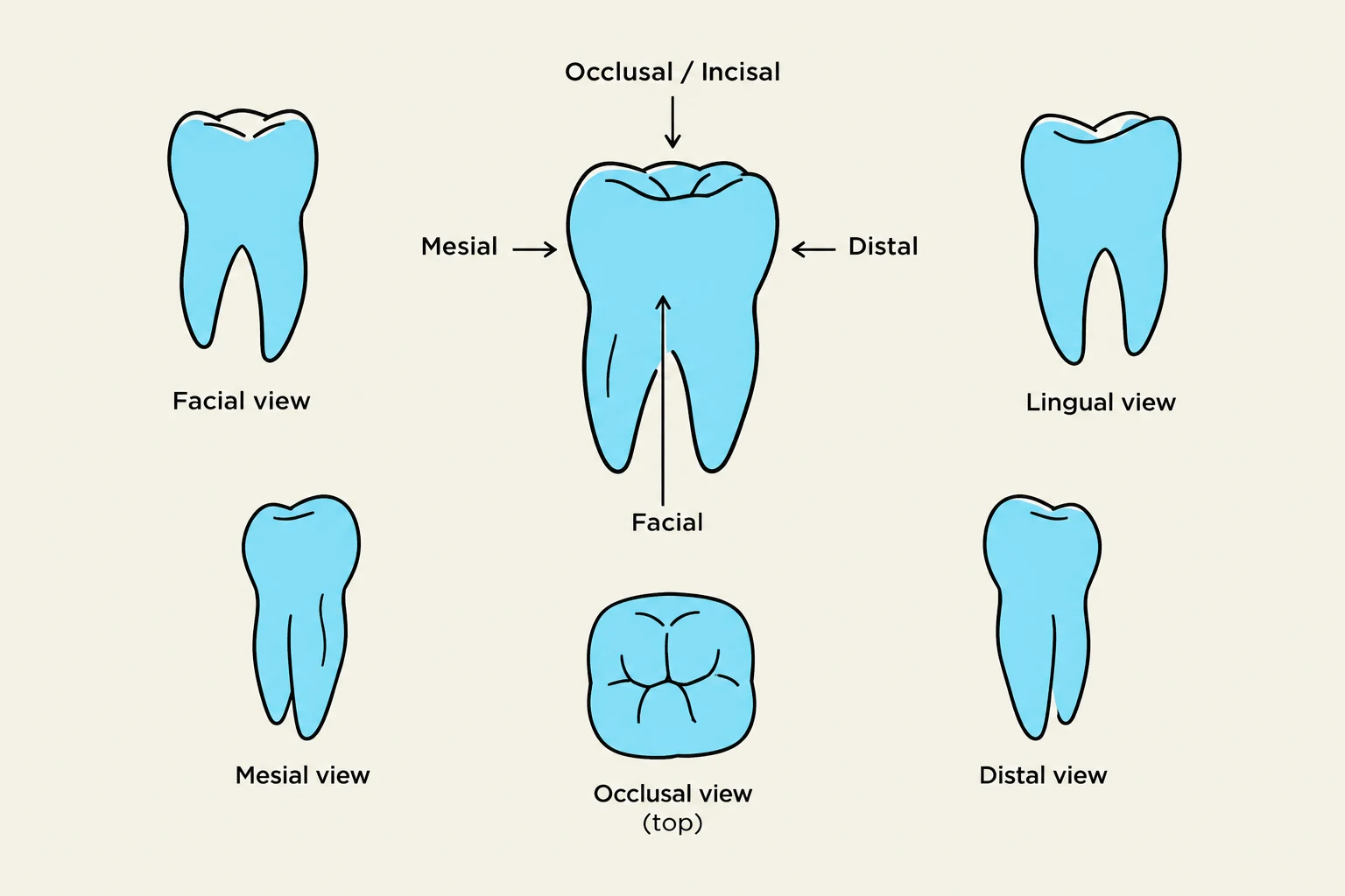
Each tooth has five primary surfaces:
Facial: Toward the face. This is subdivided into labial (for anterior teeth facing the lips) and buccal (for posterior teeth facing the cheek).
Lingual: Toward the tongue. In the maxillary arch, this may be called palatal.
Mesial: The surface closest to the midline.
Distal: The surface farthest from the midline.
Incisal/Occlusal: The masticatory (chewing) surface. Anterior teeth have an incisal edge, while posterior teeth have an occlusal surface.
The mesial and distal surfaces are collectively known as proximal surfaces.
Diagram illustrating different views of a tooth: facial, lingual, mesial, distal, and occlusal.
Crown and Root Anatomy
A tooth is divided into the crown and the root.
Anatomic vs. Clinical: The anatomic crown is the portion covered by enamel, while the clinical crown is the portion visible in the mouth. The clinical crown can change due to gingival recession or eruption.
CEJ: The cementoenamel junction (CEJ) is the external line where the enamel of the crown meets the cementum of the root.
Root Morphology: Roots taper toward the apex and may have concavities (depressions) or furcations (where roots divide).
Exam Tip: When describing a location, use the D-A-Q-T order: Dentition, Arch, Quadrant, and Tooth type/position.
Tooth Form and Function
The geometric shape of a tooth is directly related to its physiological role:
Incisors: Have a triangular proximal outline and are designed for biting and cutting.
Canines: Feature a tapered crown with a single pronounced cusp for piercing and tearing.
Premolars: Found only in permanent dentition; they act as transition teeth between canines and molars.
Molars: Possess broad occlusal tables and multiple cusps for grinding food.
Understanding these forms is essential for restorative dentistry, as any restoration must replicate the natural height of contour (the greatest convexity of a surface) to properly deflect food and protect the gingiva.
Keep reading for free.
Create an account to finish this reading and make it part of your study routine.