


When studying peripheral vascular diseases, it is critical to distinguish between arterial and venous disorders. While arterial issues often involve a lack of blood flow to the extremities, venous disorders—specifically Deep Vein Thrombosis (DVT) and thrombophlebitis—revolve around the obstruction or inflammation of the veins that return blood to the heart. Understanding the clinical presentation of these conditions is vital for early intervention and the prevention of life-threatening complications like pulmonary embolism.
Deep Vein Thrombosis (DVT)
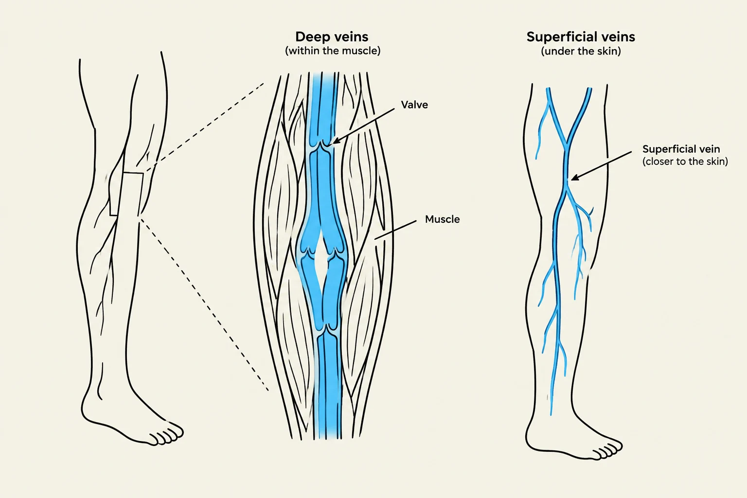
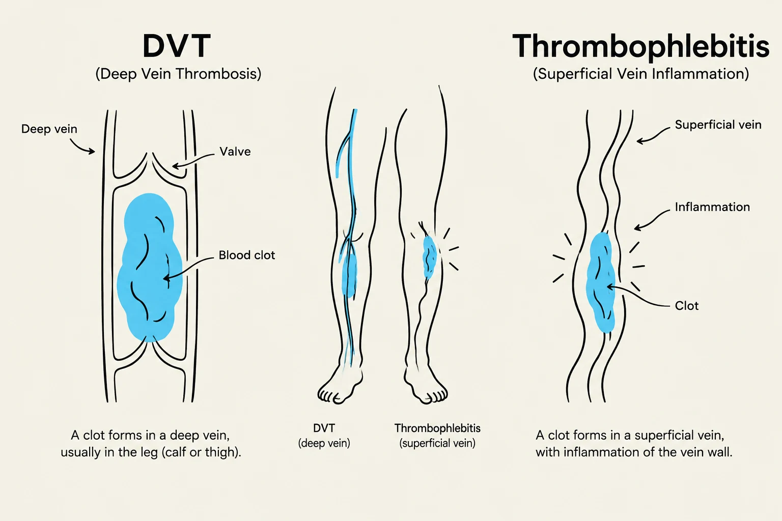
Deep Vein Thrombosis occurs when a blood clot (thrombus) forms in one or more of the deep veins in the body, most commonly in the legs. Unlike superficial veins, which are close to the skin surface, deep veins are located within the muscle layers, making them harder to palpate but much more dangerous if a clot dislodges.
The primary danger of DVT is that the clot can break loose, travel through the bloodstream, and lodge in the lungs, causing a pulmonary embolism (PE). Therefore, recognizing the early signs of localized venous obstruction is a priority in clinical practice.
Clinical Manifestations of DVT
Because a DVT obstructs the normal flow of blood back toward the heart, blood "pools" in the distal portion of the limb. This leads to a specific triad of symptoms that students should memorize:
- Localized Edema: Swelling is often unilateral (affecting only one leg). This occurs because the increased hydrostatic pressure in the veins forces fluid into the surrounding interstitial tissues.
- Warmth: The inflammatory response to the clot and the localized congestion of blood can cause the skin over the affected area to feel noticeably warmer than the surrounding skin or the opposite limb.
- Pain or Tenderness: Patients often describe a cramping sensation or localized soreness. This pain may intensify when the patient attempts to walk or flex the foot upward (dorsiflexion).
Keep reading for free.
Create an account to finish this reading and make it part of your study routine.
Continue reading freeAlready have an account? Log in
Updated