Understanding Cytoreductive Pharmacotherapy for Myeloproliferative Neoplasms
This content explains cytoreductive pharmacotherapy used in managing myeloproliferative neoplasms (MPNs). It details the clinical rationale for reducing excessive blood cell production, focusing on the risks of thrombosis and cytopenias. The document describes the mechanisms of action, clinical applications, and consid
In the management of myeloproliferative neoplasms (MPNs), the primary clinical objective is often not a complete cure, but rather the control of excessive blood cell production. When the bone marrow produces an uncontrolled surplus of red blood cells, white blood cells, or platelets, the patient faces a significantly heightened risk of life-threatening thrombotic events, such as strokes, myocardial infarctions, or deep vein thrombosis.
Cytoreductive pharmacotherapy refers to the use of specific medicinal agents designed to reduce these cellular counts to safer levels. By modulating the rate of cell production or inducing apoptosis in malignant clones, these therapies mitigate the risks of vascular occlusion and improve the overall quality of life for the patient.
The Clinical Rationale: Why Reduce Cell Counts?
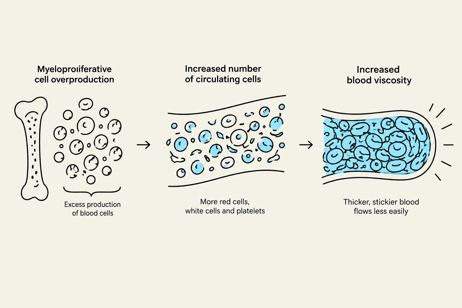
The necessity for cytoreduction is driven by the physical and rheological consequences of hypercellularity. For example, in Essential Thrombocythemia (ET) or Polycythemia Vera (PV), the sheer number of circulating cells increases blood viscosity and promotes spontaneous clot formation.
The goal of therapy is to achieve a "therapeutic window" where:
The cell counts are low enough to prevent thrombosis.
The counts are not so low that the patient develops cytopenias (deficiencies in blood cells) that lead to bleeding or infection.
Exam Tip: When evaluating a patient for cytoreduction, always assess their baseline thrombotic risk (e.g., age, history of clots) alongside their current hematocrit or platelet count. High-risk patients require more aggressive cytoreductive strategies.
This diagram illustrates the progression of myeloproliferative disorders, starting with overproduction of blood cells in the bone marrow, leading to an increased number of circulating cells, and resulting in increased blood viscosity that impedes blood flow.
Hydroxyurea: The First-Line Standard
Hydroxyurea is widely considered the cornerstone of cytoreductive therapy due to its efficacy, ease of administration, and relatively low cost.
Mechanism of Action
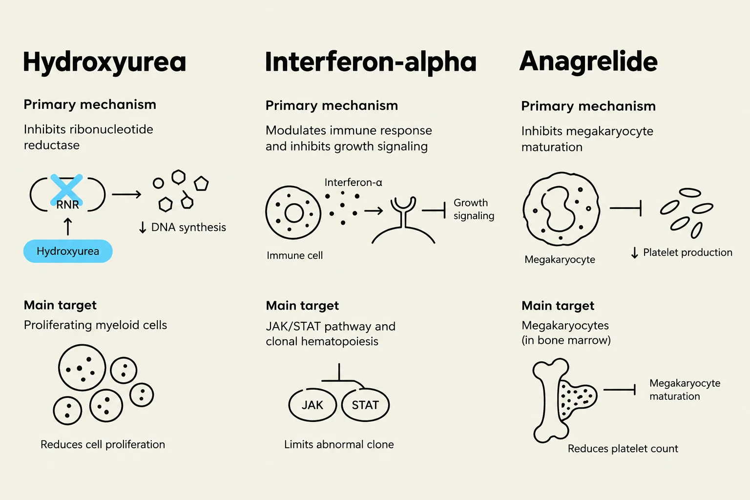
Hydroxyurea works by inhibiting the enzyme ribonucleotide reductase. This enzyme is critical for the conversion of ribonucleotides into deoxyribonucleotides, which are the essential building blocks of DNA. By limiting the availability of these building blocks, hydroxyurea effectively halts DNA synthesis during the S-phase of the cell cycle, thereby slowing down the proliferation of rapidly dividing myeloid cells.
Clinical Application
It is frequently used in:
Polycythemia Vera (PV): To reduce hematocrit levels.
Essential Thrombocythemia (ET): To lower platelet counts in high-risk patients.
Primary Myelofibrosis (PMF): To manage symptoms like splenomegaly.
While highly effective, clinicians must monitor patients closely for side effects such as myelosuppression (lowering all cell lines too much), skin ulcers, and gastrointestinal distress.
This diagram illustrates how hydroxyurea inhibits ribonucleotide reductase, leading to reduced DNA synthesis.
Interferon-alpha: An Immunomodulatory Approach
Interferon-alpha (IFN-α) represents a different pharmacological philosophy. While hydroxyurea targets the cell cycle directly, interferons act through immunomodulatory pathways.
Mechanism of Action
Interferons are cytokines that signal the body's immune system. In the context of MPNs, IFN-α is thought to:
Directly inhibit the proliferation of hematopoietic stem cells.
Induce apoptosis (programmed cell death) in the malignant clones.
Modulate the cytokine environment within the bone marrow to reduce inflammation.
Clinical Considerations
IFN-α is often preferred in specific populations, such as pregnant women (where hydroxyurea is contraindicated due to teratogenicity) or younger patients where there is a desire to potentially achieve a deeper molecular response. However, it is often associated with "flu-like" symptoms, including fever, chills, and fatigue, which can impact patient adherence.
Anagrelide: Targeted Platelet Reduction
For patients whose primary clinical concern is extreme thrombocytosis (excessive platelets) that has not responded adequately to other measures, Anagrelide provides a more specialized option.
Mechanism of Action
Unlike hydroxyurea, which affects multiple cell lines, Anagrelide is more selective for megakaryocytes (the precursor cells to platelets). It works by interfering with the maturation of these cells. Specifically, it inhibits the production of certain signaling molecules (like retinoic acid) that are necessary for megakaryocyte development, thereby reducing the number of platelets released into the bloodstream.
Clinical Profile
Target: Specifically used for Essential Thrombocythemia (ET).
Advantage: It does not typically cause the widespread suppression of red or white blood cells seen with hydroxyurea.
Risk: A significant side effect is cardiovascular, including palpitations and tachycardia (rapid heart rate), necessitating regular cardiac monitoring.
This diagram illustrates the mechanisms of action for three drugs: Hydroxyurea, Interferon-alpha, and Anagrelide, detailing their primary mechanisms, main targets, and effects on cellular processes.
Summary of Therapeutic Selection
Choosing the correct cytoreductive agent involves a complex calculation of the patient's disease subtype, their risk for thrombosis, and their ability to tolerate specific side effects.
Agent
Primary Target
Mechanism
Key Consideration
Hydroxyurea
Multiple (RBC, WBC, Platelets)
DNA synthesis inhibition
First-line; risk of myelosuppression
Interferon-α
Hematopoietic Stem Cells
Immunomodulation/Apoptosis
Preferred in pregnancy; flu-like symptoms
Anagrelide
Platelets (Megakaryocytes)
Inhibition of maturation
Selective for ET; risk of palpitations
By understanding these mechanisms, clinicians can tailor treatment to reduce the cellular burden while preserving the patient's physiological stability.
Keep reading for free.
Create an account to finish this reading and make it part of your study routine.