


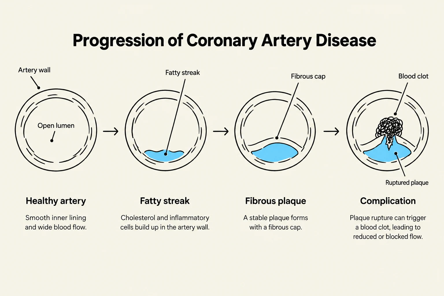
Coronary Artery Disease (CAD) is a progressive condition characterized by the narrowing or occlusion of the coronary arteries, typically due to atherosclerosis. As plaque accumulates, the blood supply to the myocardium becomes insufficient to meet metabolic demands, leading to myocardial ischemia. Understanding how to diagnose these blockages and how to pharmacologically manage the resulting symptoms and risks is essential for preventing myocardial infarction (MI) and death.
Clinical Manifestations of Ischemia
The primary clinical manifestation of CAD is angina, or chest pain, which occurs when myocardial oxygen demand exceeds the supply. It is critical to distinguish between the different patterns of angina to determine the level of urgency:
- Chronic Stable Angina: Typically precipitated by physical exertion or emotional stress.
- Unstable Angina: A more dangerous state characterized by new-onset angina that occurs at rest or with minimal exertion, often increasing in frequency, duration, or severity.
- Prinzmetal's Angina: Often caused by a coronary artery spasm rather than fixed atherosclerotic narrowing.
Exam Tip: If a patient presents with chest pain that is new, occurs at rest, or is becoming more severe, suspect unstable angina, which requires immediate medical evaluation.
Diagnostic Modalities
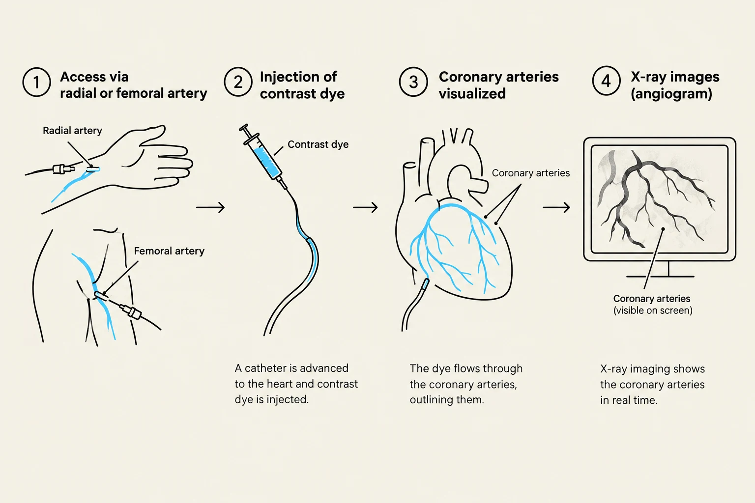
To identify the presence, location, and severity of arterial blockages, clinicians utilize a variety of non-invasive and invasive tests.
Non-Invasive Testing
The first step often involves an Electrocardiogram (12-lead EKG) to identify changes in cardiac electrical activity. It is ideal to compare the current EKG with a patient's baseline to detect new ST-segment elevations or T-wave changes.
An Echocardiogram uses ultrasound to assess cardiac function. Clinicians specifically look for left ventricular wall motion abnormalities, which can suggest areas of the heart supplied by diseased arteries. For a more detailed view, a Transesophageal Echocardiogram (TEE) may be performed, where a scope is passed down the esophagus to provide high-resolution imaging from a position closer to the heart.
Keep reading for free.
Create an account to finish this reading and make it part of your study routine.
Continue reading freeAlready have an account? Log in
Updated