


In the complex landscape of the human immune system, millions of cells circulate through the blood and tissues, each performing specialized roles. To a casual observer under a microscope, many of these cells—such as various types of white blood cells—might look remarkably similar. To distinguish a helper T cell from a cytotoxic T cell, or a mature B cell from a precursor, scientists rely on a molecular "barcode" system known as Cluster of Differentiation (CD) markers.
What are CD Markers?
Cluster of Differentiation (CD) markers are specialized proteins located on the surface of cells. These proteins, which can be receptors, adhesion molecules, or enzymes, serve as unique identifiers for specific cell types or stages of development. By using these markers, immunologists can perform immunophenotyping, a process of characterizing cells based on the specific set of proteins they express on their membranes.
Think of CD markers as a set of biological ID cards. Just as a person might be identified by a combination of height, eye color, and a fingerprint, an immune cell is identified by its "phenotype"—the specific combination of CD markers present on its surface.
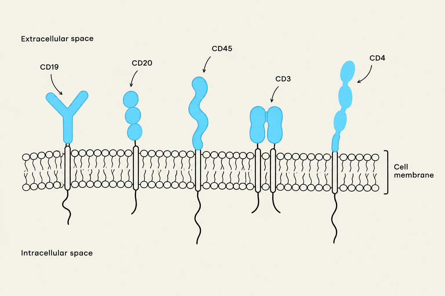
The Role of Immunophenotyping
Immunophenotyping is the practical application of CD markers. It typically involves using fluorescently labeled antibodies that are designed to bind specifically to a particular CD marker. When these antibodies are applied to a sample of cells and run through a machine called a Flow Cytometer, the machine detects the light emitted by the fluorophores.

Keep reading for free.
Create an account to finish this reading and make it part of your study routine.
Continue reading freeAlready have an account? Log in
Updated