Understanding Cardiovascular and Peripheral Vascular Disorders
A comprehensive guide to cardiovascular and peripheral vascular disorders, covering their anatomy, physiology, common conditions, diagnostic methods, and nursing management.
The cardiovascular system serves as the body's primary transport network, utilizing the heart as a pump to circulate oxygen and vital nutrients through a vast web of blood vessels. When this system is compromised, the most immediate consequence is decreased perfusion—a state where tissues do not receive enough oxygenated blood. For a nurse, recognizing the early signs of poor perfusion, such as dyspnea (shortness of breath), fatigue, confusion, or pain, is critical to preventing systemic collapse.
Anatomy, Physiology, and Electrical Conduction
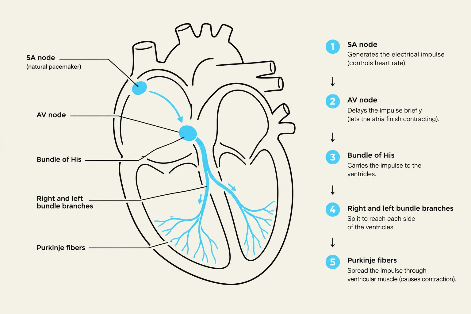
To understand cardiovascular disease, one must first understand how the heart generates and conducts the electrical impulses that drive its rhythmic contractions. The process begins at the SA node, known as the heart's natural pacemaker, which initiates the electrical impulse. This impulse travels to the AV node, which provides a brief delay to allow the ventricles to fill completely with blood before contracting. Finally, the impulse is distributed through the bundle branches and Purkinje fibers, ensuring a coordinated contraction of the ventricles.
Diagram illustrating the electrical conduction system of the heart, including the SA node, AV node, Bundle of His, bundle branches, and Purkinje fibers.
Disruptions in this electrical pathway lead to cardiac dysrhythmias. For example, atrial fibrillation is a common arrhythmia that can cause the atria to quiver rather than contract effectively, significantly increasing the risk of blood clots and subsequent stroke.
Coronary Artery Disease and Myocardial Infarction
The heart muscle itself requires a constant supply of oxygen, delivered via the coronary arteries. Coronary Artery Disease (CAD) occurs when these arteries become diseased or narrowed. The primary driver of CAD is atherosclerosis, a process characterized by the buildup of plaque within the arterial walls.
When the narrowing of these vessels limits oxygen delivery to the myocardium (heart muscle), the patient experiences angina pectoris, or chest discomfort. If the blood flow is completely blocked for a sufficient duration, the heart muscle begins to die; this clinical event is known as a Myocardial Infarction (MI).
Exam Tip: When assessing for an MI, remember that symptoms are not always "classic." While many experience chest pressure, women may present with atypical symptoms rather than acute chest pain.
Cardiac Diagnostics
To differentiate between stable angina and an acute MI, clinicians rely on specific diagnostic markers:
Troponin I: A protein released into the bloodstream specifically when myocardial damage has occurred.
BNP (B-type Natriuretic Peptide): A marker that rises when the ventricles are under pressure overload, often indicating heart failure.
EKG/ECG: Used to monitor the electrical activity and identify rhythm changes or signs of ischemia.
Heart Failure and Hypertension
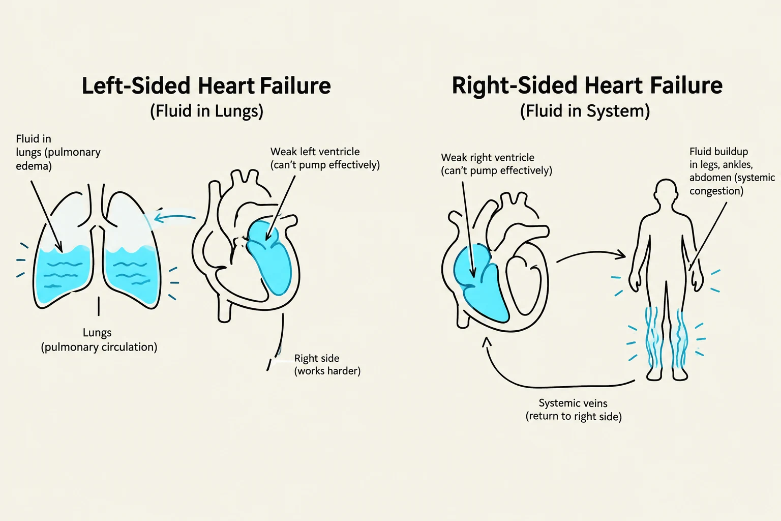
Heart failure is a chronic condition where the heart is unable to pump enough blood to meet the metabolic demands of the body. It is classified based on where the "backup" of fluid occurs:
Left-sided failure: The pump fails on the left side, causing blood to back up into the lungs, leading to respiratory symptoms.
Right-sided failure: The pump fails on the right side, causing blood to back up into the systemic circulation (the body), leading to peripheral edema.
Diagram illustrating the differences between left-sided and right-sided heart failure, showing fluid accumulation in the lungs for left-sided failure and systemic congestion in the body for right-sided failure.
Parallel to these issues is hypertension (high blood pressure). It is categorized into three types:
Essential: No single identifiable cause.
Secondary: Caused by an underlying disorder.
Malignant: A severe, rapid rise in blood pressure.
Because hypertension is often asymptomatic, it is frequently called a "silent killer." If left uncontrolled, it can lead to significant damage to target organs like the heart, kidneys, and brain.
Peripheral Vascular Disorders (PVD)
Peripheral Vascular Disorders is an umbrella term that includes both arterial and venous issues. Understanding the difference between "flow to the tissue" and "return from the tissue" is essential for nursing assessment.
Peripheral Artery Disease (PAD)
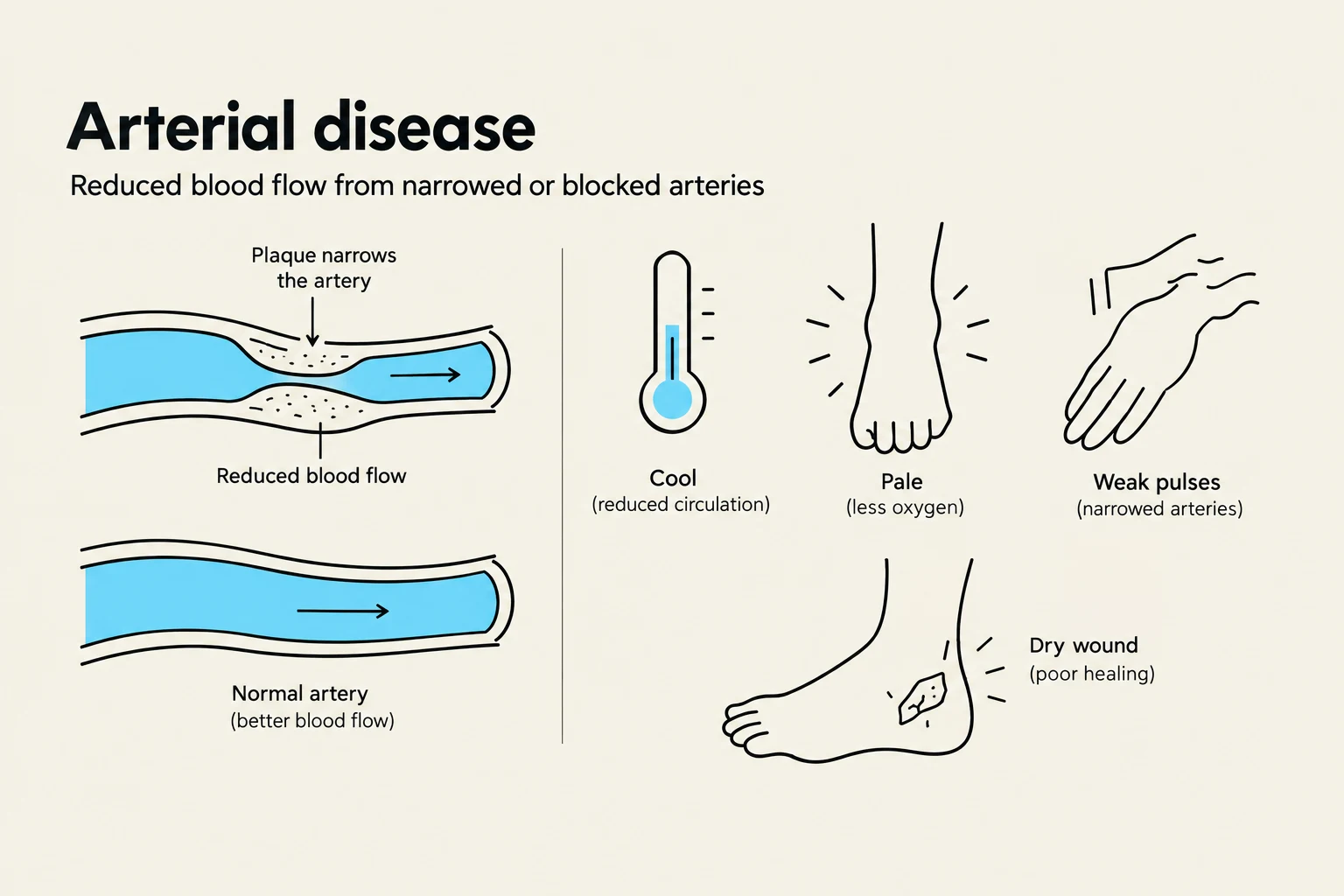
PAD involves a reduction in blood flow from the heart to the peripheral tissues. Because the tissues are starved of oxygen (ischemia), patients often present with:
Cool, pale skin.
Weak or absent pulses.
Ischemic pain during activity (claudication) that is relieved by rest.
Dry wounds, often on the toes.
Venous Disorders
Venous disorders involve poor blood return back to the heart, causing blood to pool in the extremities. This pooling leads to:
Edema (swelling) and warmth.
Discoloration of the skin.
Varicose veins and thrombophlebitis (inflammation of a vein due to a clot).
Stasis ulcers: Moist wounds typically found around the ankles due to chronic swelling.
Diagram illustrating arterial disease, showing plaque narrowing an artery and its symptoms such as reduced blood flow, coolness, paleness, weak pulses, and dry wounds.
Nursing Management and Priorities
Nursing care for cardiovascular patients focuses on three pillars: assessment, intervention, and education.
Clinical Interventions:
Oxygenation and Circulation: Monitor vital signs closely and support oxygenation to ensure tissues are perfused.
Fluid Management: In heart failure patients, monitor for electrolyte imbalances and be cautious with rapid IV fluid administration, as this can worsen congestion.
Safety: Monitor for dysrhythmias and report any changes in cardiac rhythm or pulse quality immediately.
Patient Education:
Effective management often requires lifestyle modifications to reduce risk factors such as smoking, obesity, and hyperlipidemia.
For PAD: Advise patients to stop smoking, keep legs warm, and avoid tight clothing.
For Venous Disorders: Teach patients to elevate their legs, avoid prolonged standing, and use compression stockings if prescribed.
For Heart Failure: Educate on low-sodium diets, fluid restrictions, and the importance of daily weight monitoring to detect fluid retention early.
Keep reading for free.
Create an account to finish this reading and make it part of your study routine.