


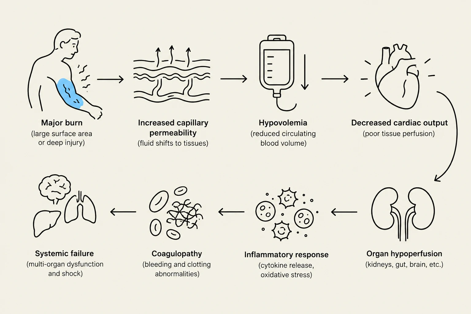
Burn injuries are complex traumatic events that can affect nearly every physiological system in the human body. Beyond the visible damage to the skin, burns trigger massive inflammatory responses, fluid shifts, and systemic metabolic changes. Understanding how to classify these injuries by their depth and mechanism is the first step in effective clinical management and triage.
Classification by Depth of Tissue Damage
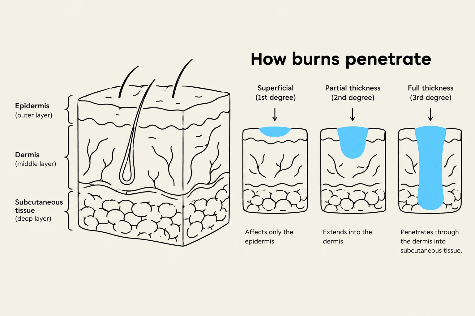
Clinicians categorize burns based on how deeply the injury penetrates the skin layers. This classification is vital because it dictates the healing time, the risk of infection, and the necessity for surgical intervention.
Superficial (First-degree) Burns
These burns affect only the epidermal layer. Common causes include severe sunburns, brief flash burns, or minor splashes of hot liquid. These injuries are typically characterized by redness and pain but usually heal within a week without leaving permanent scars.
Partial-thickness (Second-degree) Burns
Partial-thickness burns involve the epidermis and varying depths of the dermal layer. They can be subdivided into superficial or deep partial-thickness depending on the extent of tissue necrosis. Key clinical features include:
- Edema (swelling) and the formation of epidermal blisters.
- A wet, raw, pink, or cherry-red appearance that blanches when pressure is applied.
- High levels of pain due to exposed nerve endings.
These burns may heal within three to six weeks, but they often result in some degree of scarring or changes in skin pigmentation.
Full-thickness (Third-degree) Burns
Full-thickness burns are the most severe, destroying the epidermis, dermis, hair follicles, and all underlying structures. The skin may appear white, brown, black, or red, and the surrounding tissue is often heavily edematous.
Keep reading for free.
Create an account to finish this reading and make it part of your study routine.
Continue reading freeAlready have an account? Log in
Updated