


The lungs are complex organs responsible for gas exchange, a vital process for life. When this process is disrupted, it can lead to serious conditions like atelectasis and pulmonary edema. Atelectasis refers to the collapse of alveoli, the tiny air sacs in the lungs, while pulmonary edema involves the accumulation of fluid in the lung tissue. Understanding the mechanisms behind these conditions is crucial for diagnosis and treatment.
Atelectasis: The Collapse of Alveoli
Atelectasis occurs when alveoli, the primary sites of gas exchange, collapse and are unable to participate in respiration. This collapse can be caused by several factors: external pressure on the lungs, such as from obesity or a tumor; decreased ventilation, often seen in post-operative patients who breathe shallowly due to pain; or a reduction in surfactant. Surfactant is a substance that reduces surface tension within the alveoli, preventing them from collapsing. Premature infants are particularly at risk due to insufficient surfactant production.
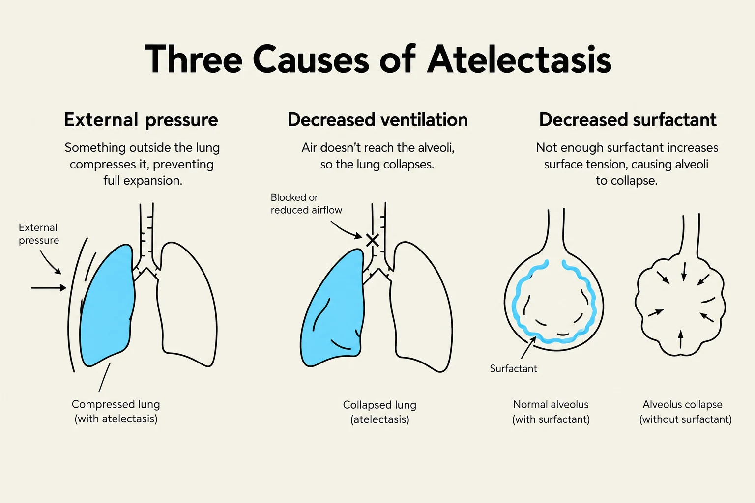
Symptoms of atelectasis can include shortness of breath and hypoxemia (low oxygen levels in the blood). Diagnosis can be challenging as imaging techniques may not readily detect the collapse of individual alveoli. Pulmonary function tests (PFTs) are often used to assess lung capacity and airflow. Treatment focuses on addressing the underlying cause and encouraging deep breathing. Devices like incentive spirometers help patients take deeper breaths, promoting alveolar expansion. Positive pressure ventilation may also be used to help keep alveoli open.
Keep reading for free.
Create an account to finish this reading and make it part of your study routine.
Continue reading freeAlready have an account? Log in
Updated