


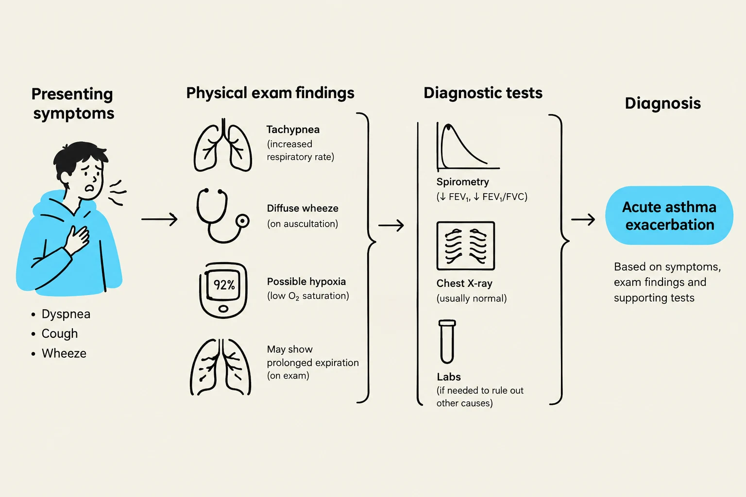
In clinical practice, respiratory assessments require a systematic approach to differentiate between various obstructive and infectious pathologies. This article examines the case of JJ, a 14-year-old female presenting with acute respiratory distress, to illustrate the diagnostic process and the physical examination techniques used to identify an asthma exacerbation.
Patient Presentation and Clinical History
JJ presented to urgent care with a sudden onset of severe respiratory symptoms that had progressed over the course of one hour. She reported a sensation of "breathing through a straw," which she rated as a 9/10 in severity. Her primary complaints included:
- Shortness of breath (dyspnea): A common chief concern in respiratory assessments.
- Chest tightness: A generalized sensation of pressure in the thoracic region.
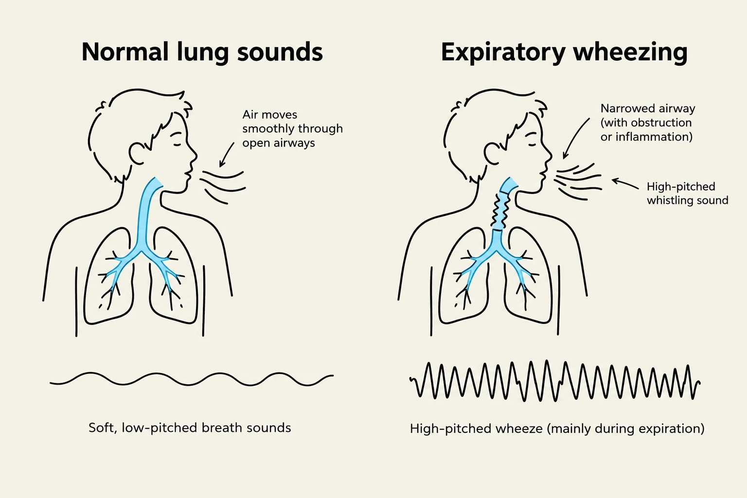
- Wheezing and Coughing: A nonproductive cough had been present since the previous night, followed by audible wheezing in the morning.
The patient's medical history is significant for asthma diagnosed in early childhood, which was triggered by environmental factors such as wood-burning smoke. In this acute episode, the suspected trigger was secondhand cigarette smoke exposure while staying at a friend's house. JJ typically uses an albuterol inhaler as needed (prn) for exercise-induced symptoms, but she did not have her medication available during this episode.
Clinical Note: When assessing a patient with respiratory distress, always observe for "red flags" such as labored breathing, cyanosis (bluish discoloration), or the use of accessory muscles to breathe, as these indicate acute respiratory distress.
Physical Examination and Vital Signs
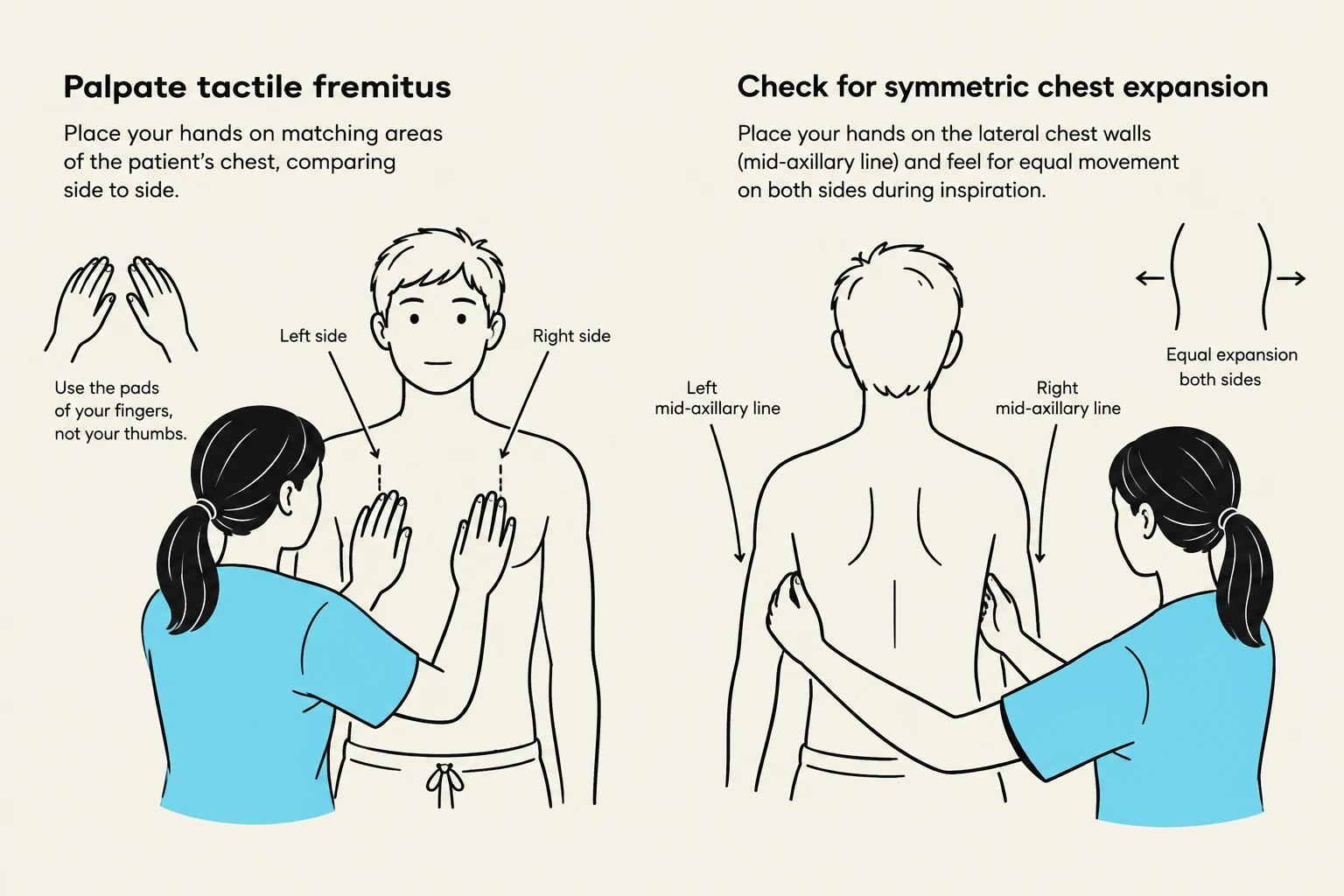
A thorough physical examination is essential to quantify the severity of the exacerbation and guide treatment.
Keep reading for free.
Create an account to finish this reading and make it part of your study routine.
Continue reading freeAlready have an account? Log in
Updated