Understanding Asthma and COPD: Pathophysiology and Nursing Management
This article provides a comprehensive overview of the respiratory system, focusing on the pathophysiology, assessment, and nursing management of asthma and Chronic Obstructive Pulmonary Disease (COPD). It covers fundamental respiratory function, age-related changes, and the clinical manifestations of these conditions.
Understanding respiratory health requires a grasp of how the body moves oxygen into the bloodstream and removes carbon dioxide. When these processes are disrupted by chronic conditions like asthma or Chronic Obstructive Pulmonary Disease (COPD), patients face significant challenges in gas exchange and perfusion. This article explores the pathophysiology, assessment, and management of these critical respiratory disorders.
Fundamentals of Respiratory Function
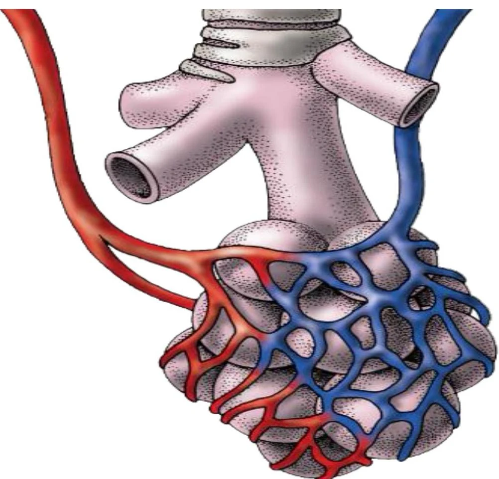
The primary goal of the respiratory system is efficient gas exchange—the movement of oxygen into the blood and carbon dioxide out of it. This process is inextricably linked to perfusion, which is the movement of blood through the pulmonary circulation to facilitate this exchange.
Illustration of the pulmonary alveoli and capillaries, showing the exchange of oxygen and carbon dioxide.
Age-Related and Environmental Changes
As humans age, several physiological changes occur that can impact respiratory efficiency:
Decreased lung volume and elasticity: The lungs become less efficient at expanding and contracting.
Decreased alveolar surface area: This reduces the space available for gas exchange.
Reduced muscle strength: Weakness in the diaphragm and intercostal muscles makes breathing more labored.
Decreased cough reflex: This makes it harder for the body to clear unwanted particles or secretions.
Furthermore, immobility poses a significant risk to respiratory health. Lack of movement can lead to the pooling of thick secretions, a decreased ability to cough effectively, and an increased risk of atelectasis (the partial or complete collapse of a lung lobe) and pneumonia.
Asthma: Airway Hyperresponsiveness
Asthma is a condition characterized by intermittent and reversible airflow obstruction. It is driven by airway hyperresponsiveness and inflammation.
In an untreated asthmatic airway, several pathological changes occur: the airways become narrowed, the muscles surrounding the airways undergo bronchoconstriction, and the airway walls become inflamed and damaged. This often manifests clinically as chest tightness, wheezing, dyspnea (shortness of breath), and coughing.
Nursing Tip: Management of asthma focuses on both "control" therapy (daily medications to prevent attacks) and "reliever" therapy (medications like bronchodilators to stop an acute attack in progress).
COPD: Chronic Obstructive Pulmonary Disease
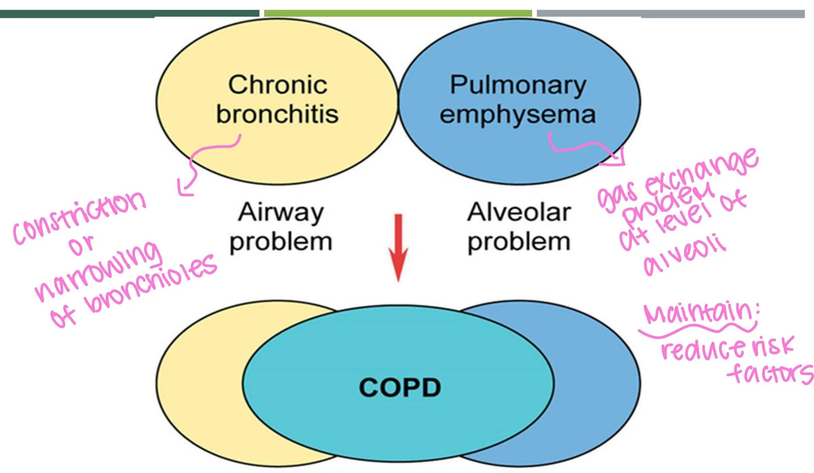
COPD is an "umbrella" term for chronic airflow limitation, most commonly encompassing chronic bronchitis and emphysema. It is the fourth leading cause of mortality in the U.S., with cigarette smoking being the greatest risk factor.
Chronic Bronchitis vs. Emphysema
While both contribute to COPD, they affect different parts of the respiratory anatomy:
Chronic Bronchitis: This is primarily an airway problem. Chronic exposure to irritants causes inflammation of the bronchi and bronchioles, leading to mucosal edema and the production of large amounts of thick mucus.
Emphysema: This is primarily an alveolar problem. High protease levels cause alveolar damage and the breakdown of elastin, leading to a loss of lung elasticity. This results in air trapping and hyperinflation of the lungs.
This diagram illustrates the relationship between chronic bronchitis and pulmonary emphysema, both of which are airway and alveolar problems respectively, and contribute to COPD. It also highlights the importance of reducing risk factors for maintenance.
Clinical Manifestations of COPD
Patients with advanced COPD often present with distinct physical findings:
Barrel Chest: Caused by chronic hyperinflation and air trapping.
Cyanosis: A bluish discoloration of the skin or mucosa, indicating poor perfusion and low oxygen levels.
Hypoxemia: Low levels of oxygen in the blood.
Digital Clubbing: Bulbous enlargement of the fingertips.
Cor Pulmonale: Right-sided heart failure resulting from long-term pulmonary disease.
Assessment and Diagnostics
A focused respiratory assessment is vital for identifying alterations in function. Nurses should collect a history regarding work environments and smoking habits, and perform physical assessments including inspection for chest symmetry and percussion.
Laboratory and Diagnostic Tools
Arterial Blood Gases (ABGs): Used to assess oxygenation (PaO2), ventilation (PaCO2), and acid-base status (HCO3).
Note: Hypoventilation can lead to respiratory acidosis, characterized by a low pH and high PaCO2.
Pulmonary Function Tests (PFTs): Measure vital capacity, residual volume, and total lung capacity.
Chest X-ray: Can reveal hyperinflated lungs in COPD patients.
Sputum Culture and CBC: To check for infections (e.g., elevated WBC) or polycythemia (excessive RBCs due to chronic low oxygen).
Nursing Management and Interventions
Management of respiratory disease is holistic, focusing on improving gas exchange, reducing anxiety, and preventing infection.
Non-Surgical Interventions
Breathing Techniques:
Pursed-lip breathing: Helps maintain airway pressure to prevent airway collapse.
Diaphragmatic breathing: Focuses on using the diaphragm rather than accessory muscles.
Positioning: The tripod position (leaning forward) can help ease the work of breathing.
Incentive Spirometry: Used to promote lung expansion, especially post-surgery.
Oxygen Therapy
Oxygen must be administered with caution, as excessive oxygen in some patients can depress respirations.
Nasal Cannula: For low-level oxygen needs (2-6 L/min).
Simple Mask: For moderate oxygen needs (5-10 L/min).
Venturi Mask: Delivers the most accurate, constant concentration of oxygen; ideal for chronic lung disorders.
Non-rebreather Mask: Used in emergencies to provide high concentrations (up to 100%).
CPAP/BiPAP: Continuous or Bilevel Positive Airway Pressure devices help keep alveoli open and assist with ventilation.
Pharmacological Interventions
Medications are categorized by their function:
Adrenergics (e.g., Albuterol): Bronchodilators to open airways.
Anticholinergics (e.g., Ipratropium): To reduce bronchoconstriction.
Corticosteroids (e.g., Prednisone): To reduce inflammation.
Mucolytics (e.g., Acetylcysteine): To thin thick secretions.
Dietary and Lifestyle Support
Patients with COPD often struggle with weight loss due to the high energy cost of breathing. A diet that is high calorie, high protein, and high fat, but low carbohydrate and low sodium, is often recommended. Additionally, smoking cessation is the most critical intervention to prevent further lung damage.
Exam Tip: When managing COPD, remember that "small, frequent meals" and "increasing fluids to thin secretions" are key nursing interventions to support nutrition and airway clearance.
Keep reading for free.
Create an account to finish this reading and make it part of your study routine.