Understanding Anti-Nuclear Antibody (ANA) Tests for Autoimmune Diseases
This guide explains the principles and interpretation of Anti-Nuclear Antibody (ANA) tests, a crucial tool in diagnosing autoimmune diseases. It details the Indirect Immunofluorescence (IFA) screening method, common ANA patterns (homogeneous, rim, speckled, nucleolar, centromere), and confirmatory testing for specific
The diagnosis of autoimmune diseases often begins with a screening process designed to detect antibodies that target the body's own cellular components. One of the most critical tools in this process is the Anti-Nuclear Antibody (ANA) test. By identifying antibodies that react against antigens located within the cell nucleus, clinicians can gain significant insight into a patient's immunological status and monitor the progression of various systemic diseases.
The Fundamentals of ANA Screening
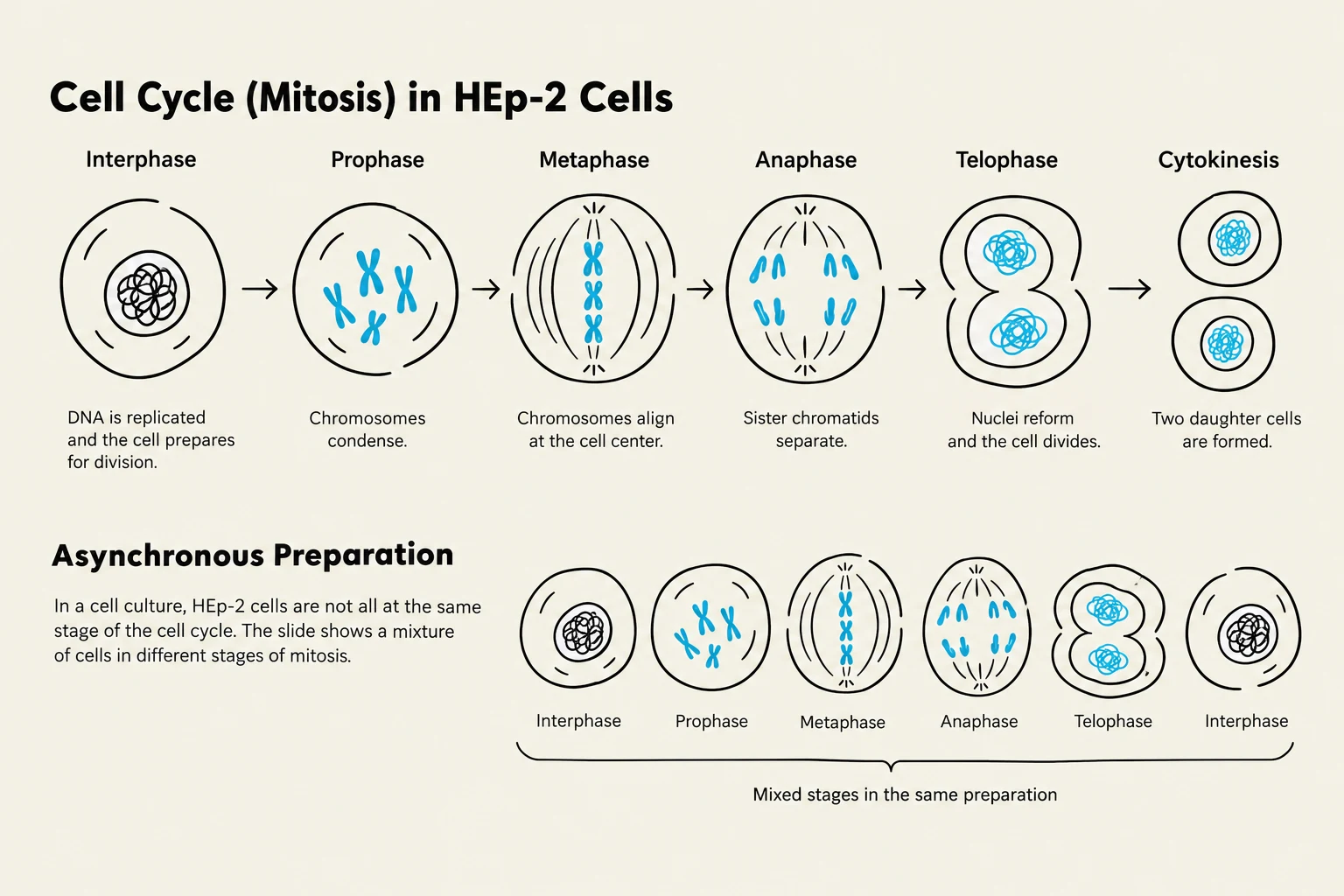
The standard method for ANA screening is Indirect Immunofluorescence (IFA) using the HEp-2 cell line. The HEp-2 cell line consists of human epithelial cells that are highly valued in the laboratory because of their large nuclei and high rate of mitosis (cell division).
To ensure a comprehensive view of the various antigens present, laboratories use an "asynchronous preparation," meaning the cells are in different stages of the cell cycle during testing. This allows for the detection of antibodies that may target different structures, such as chromatin during metaphase or nucleolar proteins during interphase.
Clinical Note on Titers: While low levels of ANA can be found in healthy individuals (especially the elderly), a titer of $1:160$ or greater is generally considered clinically significant. Note that reference ranges may vary between different laboratories.
Diagram illustrating the stages of the cell cycle (mitosis) in HEp-2 cells, including Interphase, Prophase, Metaphase, Anaphase, Telophase, and Cytokinesis. The lower section shows asynchronous preparation with a mixture of cells in different mitotic stages.
Patterns of Identity: Interpreting IFA Results
When an ANA test is positive, the next step is to observe the "pattern" of fluorescence under a microscope. These patterns provide clues about which specific antigens the antibodies are attacking.
1. Homogeneous Pattern
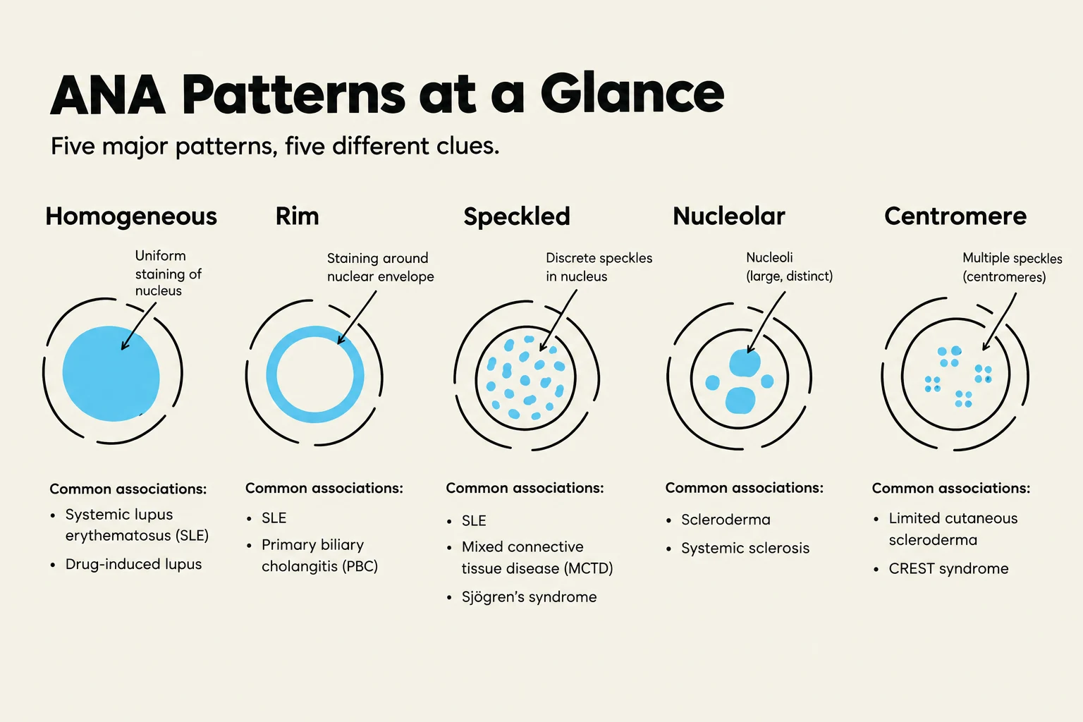
The homogeneous pattern is the most common observation. It is characterized by uniform staining of the entire nucleus. During metaphase, the staining is concentrated in the chromosome region. This pattern is typically associated with antibodies against DNA, DNP (deoxyribose nucleoprotein), and histones. It is frequently seen in Systemic Lupus Erythematosus (SLE), Sjögren syndrome, Rheumatoid Arthritis (RA), and Mixed Connective Tissue Disease (MCTD).
2. Rim / Peripheral Pattern
The rim pattern presents as solid staining around the outer region of the nucleus. Like the homogeneous pattern, it involves antigens such as DNA, DNP, and histones. This pattern is highly suggestive of active SLE.
3. Speckled Pattern
The speckled pattern consists of discrete speckles of varying sizes throughout the nucleus. In metaphase cells, these speckles appear in the non-chromosome regions. This pattern is associated with Extractable Nuclear Antigens (ENAs), including Smith (Sm), RNP, SSA/SSB, and Scl-70. It is seen in a wide range of diseases, including SLE, Sjögren syndrome, RA, MCTD, and progressive systemic sclerosis.
4. Nucleolar Pattern
The nucleolar pattern is characterized by large, coarse speckled staining, usually appearing in fewer than six spots per cell. This pattern targets antigens such as Scl-70, RNA polymerase, and nucleolar proteins. It is most commonly associated with Sjögren syndrome and progressive systemic sclerosis.
5. Centromere Pattern
The centromere pattern is unique because it presents as discrete nuclear speckles that are often multiples of 23. This is because the antibodies target the centromere chromatin of the 23 pairs of chromosomes. This pattern is highly selective for the CREST variant of progressive systemic sclerosis, which is considered a milder form of the disease.
This chart illustrates five common patterns of antinuclear antibodies (ANA) and their associated conditions, including Homogeneous, Rim, Speckled, Nucleolar, and Centromere patterns.
Confirmatory Testing and Specific Antibodies
If an ANA screening test is positive, clinicians must perform confirmatory testing to identify the specific antibodies present. This moves the diagnosis from a general "autoimmune" state to a specific disease.
Anti-double stranded DNA (dsDNA)
Anti-dsDNA antibodies (also known as native DNA) are highly specific for SLE. Their presence is considered diagnostic, and their levels often correlate with disease activity. To confirm these, the Crithidia test is used. This test utilizes C. luciliae, a protozoan containing a kinetoplast with "bare" dsDNA that is not contaminated with histone antigens.
Extractable Nuclear Antigens (ENAs)
ENAs are saline-extractable antigens, such as ribonucleoproteins and non-histone proteins. They are often named after the patients or the diseases they characterize:
Anti-Smith (Anti-Sm): Highly specific for SLE, found in approximately 30% of patients.
Anti-SSA/Ro and Anti-SSB/La: Primarily associated with Sjögren syndrome.
Anti-RNP: Found in both SLE and Mixed Connective Tissue Disease (MCTD).
Anti-Scl-70 (Anti-topoisomerase I): A marker for progressive systemic sclerosis (scleroderma) that indicates a poor prognosis.
Anti-histone Antibodies
Anti-histone antibodies are often associated with drug-induced SLE. If these antibodies are present in the absence of other specific autoantibodies, it is highly likely the patient is experiencing lupus due to medication.
Exam Tip: If you see a question regarding a patient with a "poor prognosis" in scleroderma, look specifically for the Anti-Scl-70 antibody.
Summary of Clinical Correlations
The following table summarizes the relationship between patterns, antigens, and diseases:
Pattern
Primary Antigens
Associated Diseases
Homogeneous
DNA, DNP, Histone
SLE, Sjögren, RA, MCTD
Rim
DNA, DNP, Histone
Active SLE
Speckled
Smith, RNP, SSA/SSB, Scl-70
SLE, Sjögren, RA, MCTD, Scleroderma
Nucleolar
RNA polymerase, Scl-70
Sjögren, Scleroderma
Centromere
Centromere chromatin
CREST (Scleroderma variant)
Keep reading for free.
Create an account to finish this reading and make it part of your study routine.