Understanding and Managing Vascular and Cerebrovascular Disorders
An overview of peripheral vascular diseases (arterial and venous), cerebrovascular accidents (strokes), their causes, clinical manifestations, and management strategies, including lifestyle modifications, pharmacological treatments, and surgical interventions.
Vascular disorders represent a significant challenge to patient health, primarily because they disrupt perfusion—the vital passage of oxygenated capillary blood through body tissues. For perfusion to be adequate, both the respiratory and cardiac systems must function in harmony. When this process is interrupted, whether in the extremities or the brain, it can lead to tissue hypoxia, permanent damage, or death. This article explores the pathophysiology, clinical manifestations, and management strategies for peripheral arterial disease, peripheral venous disease, and cerebrovascular accidents.
Peripheral Vascular Disease (PVD)
Peripheral vascular disease is a slow and progressive circulation disorder that affects the arteries and veins of the peripheral circulation, most commonly in the lower extremities. While the terms Peripheral Arterial Disease (PAD) and Peripheral Venous Disease (PVD) are often used interchangeably, they represent two distinct pathophysiological processes.
Peripheral Arterial Disease (PAD)
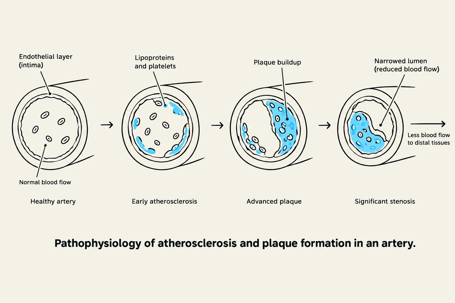
This diagram illustrates the pathophysiology of atherosclerosis and plaque formation in an artery, showing the progression from a healthy artery to significant stenosis.
PAD is a chronic condition characterized by systemic atherosclerosis. This process begins when mechanical or chemical injury occurs to the endothelial cells, altering the intima (the inner lining of the artery). This damage makes the area prone to the accumulation of platelets, lipoproteins, and debris, which eventually forms plaque. As the plaque grows, it narrows the arterial lumen, leading to tissue hypoxia distal to the obstruction.
Clinical Manifestations of PAD
The severity of PAD is often categorized into stages based on symptoms:
Stage I (Asymptomatic): No claudication is present, though pedal pulses may be weak or absent.
Stage II (Claudication): Muscle pain, cramping, or burning that occurs during exercise and is relieved by rest.
Stage III (Rest Pain): Severe, numbing, or burning pain that occurs while resting and may awaken the patient at night.
Stage IV (Necrosis/Gangrene): The presence of ulcers or blackened, gangrenous tissue, often accompanied by a distinctive odor.
Associated Arterial Conditions
Buerger’s Disease (Thromboangiitis Obliterans): An inflammatory condition that leads to the formation of clots in small to medium-sized arteries.
Raynaud’s Phenomenon: A vasospastic disorder where the smallest arteries in the fingers and toes constrict in response to cold or emotional stress, causing a "white-blue-red" color change.
Peripheral Venous Disease (PVD)
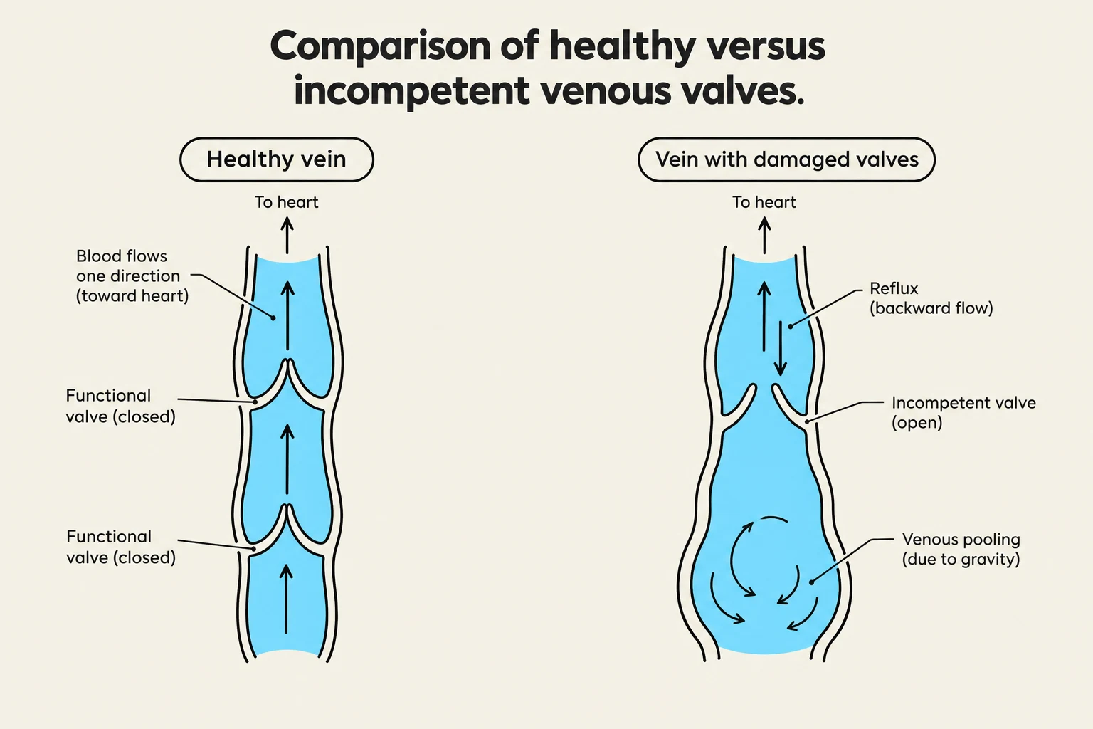
Comparison of healthy versus incompetent venous valves, illustrating blood flow and valve function.
Unlike arterial disease, which is a problem of "supply," venous disease is often a problem of "return." It involves the failure of blood to return from the extremities to the heart.
Chronic Venous Insufficiency: Occurs when one or more veins fail to return blood adequately due to damaged venous valves.
Deep Vein Thrombosis (DVT): A clot in a deep vein that carries the significant risk of traveling to the lungs (pulmonary embolism).
Thrombophlebitis: Inflammation of a vein accompanied by a clot, which can be either superficial or deep.
Varicose Veins: Dilated, twisted veins that result from incompetent valves.
Clinical Comparison: Arterial vs. Venous Ulcers
Feature
Arterial Ulcers
Venous Ulcers
Prevalence
~20% of ulcers
~75% of leg ulcers
Location
Tips of toes, webs, upper foot
Above the medial malleolus
Appearance
Small, deep, circular, pale bed
Superficial, pink bed, uneven edges
Pain Level
Very painful
Minimal pain
Other Signs
Little granulation tissue
Edema, brown discoloration
Management of Arterial Disease
Management focuses on lifestyle modification, pharmacological intervention, and, if necessary, surgical revascularization.
Lifestyle and Risk Management
To prevent and manage PAD, patients should focus on:
Smoking Cessation: This is critical; nicotine is a major risk factor.
Exercise: Gradual walking to the point of claudication, resting, and then resuming can help build collateral circulation.
Diet and Weight Control: Managing diabetes, hypertension, and hyperlipidemia through a healthy diet (low saturated fat, high fiber).
Pharmacological Therapies
Antiplatelet Agents: Drugs like Aspirin and Clopidogrel prevent platelets from sticking together.
Statins: (e.g., Atorvastatin) Lower LDL cholesterol by blocking the HMG-CoA reductase enzyme.
Phosphodiesterase Inhibitors: (e.g., Cilostazol) Used for disabling intermittent claudication due to its vasodilator properties.
Surgical Interventions
If symptoms are progressive or gangrenous, revascularization may be required:
Nonsurgical: Percutaneous Transluminal Angioplasty (PTA) using balloons or stents, or Atherectomy to mechanically remove plaque.
Surgical Bypass: A Femoral-Popliteal Bypass uses a graft (autologous or synthetic) to reroute blood around the occlusion.
Amputation: A last resort when circulation cannot be restored. Nursing care must address both physical healing and the significant psychosocial impact of limb loss.
Cerebrovascular Accident (CVA/Stroke)
A stroke is a "brain attack" resulting from a disruption in cerebral perfusion. Approximately 795,000 people experience a stroke each year.
Types of Stroke
Comparison of ischemic and hemorrhagic stroke mechanisms, illustrating blocked arteries and ruptured vessels in the brain.
Ischemic Stroke: Caused by an interruption of blood flow, often due to a clot (thrombotic or embolic) or unknown causes (cryptogenic).
Transient Ischemic Attack (TIA): A temporary loss of neurological function lasting less than 24 hours; often a warning sign of a future stroke.
Hemorrhagic Stroke: Caused by bleeding within or around the brain (e.g., intracranial aneurysm or arteriovenous malformation).
Clinical Manifestations and Assessment
Ischemic strokes often occur in the early morning, while hemorrhagic strokes may occur during physical activity. Common signs include:
Motor/Sensory Loss: Hemiplegia (paralysis), hemiparesis (weakness), or ataxia (unsteady gait).
Communication Deficits: Aphasia (expressive, receptive, or global) and dysarthria (difficulty speaking due to muscle paralysis).
Perceptual Disturbances: Hemianopsia (blindness in half the visual field) or diplopia (double vision).
Exam Tip: The ABCD Assessment Tool
Use this to determine stroke risk:
Age: Over 60 increases risk.
Blood Pressure: Greater than 140/90 mmHg.
Clinical TIA features: One-sided weakness.
Duration of symptoms: Longer duration indicates higher risk.
Medical Management of Ischemic Stroke
For patients within the appropriate window (usually 4.5 hours from onset), t-PA (tissue Plasminogen Activator) can be administered to dissolve the clot.
t-PA Inclusion/Exclusion Criteria:
Include: Age ≥ 18, clinical diagnosis of ischemic stroke, onset ≤ 4.5 hours, and BP < 185/110 mmHg.
Exclude: Recent head trauma/surgery, active internal bleeding, or history of both stroke and diabetes.
For those not receiving t-PA, management includes anticoagulants (Heparin, Warfarin), maintaining cerebral hemodynamics (elevating the head of the bed to 30 degrees), and monitoring for complications like increased intracranial pressure.
Nursing Management of Stroke
Nursing care focuses on optimizing cerebral perfusion and preventing complications:
Aneurysm Precautions: For hemorrhagic risks.
Rehabilitation: Assisting with mobility, nutrition, and communication.
Core Measures: Ensuring patients are discharged on appropriate antithrombotic and statin therapies.
Keep reading for free.
Create an account to finish this reading and make it part of your study routine.