


Molluscum contagiosum is a common, benign skin condition caused by a member of the poxvirus family. While it is generally not a medical emergency, its highly contagious nature and distinctive physical appearance often cause significant concern for patients. For healthcare students and clinicians, recognizing the hallmark lesions and understanding the nuances of treatment—balancing patience with intervention—is essential for effective patient education and management.
Pathophysiology and Transmission
The condition is driven by the Molluscum contagiosum virus (MCV), a large, double-stranded DNA virus belonging to the Poxviridae family. Unlike many other viruses that replicate deep within the systemic circulation, MCV is primarily localized to the epidermis, where it causes the proliferation of keratinocytes.
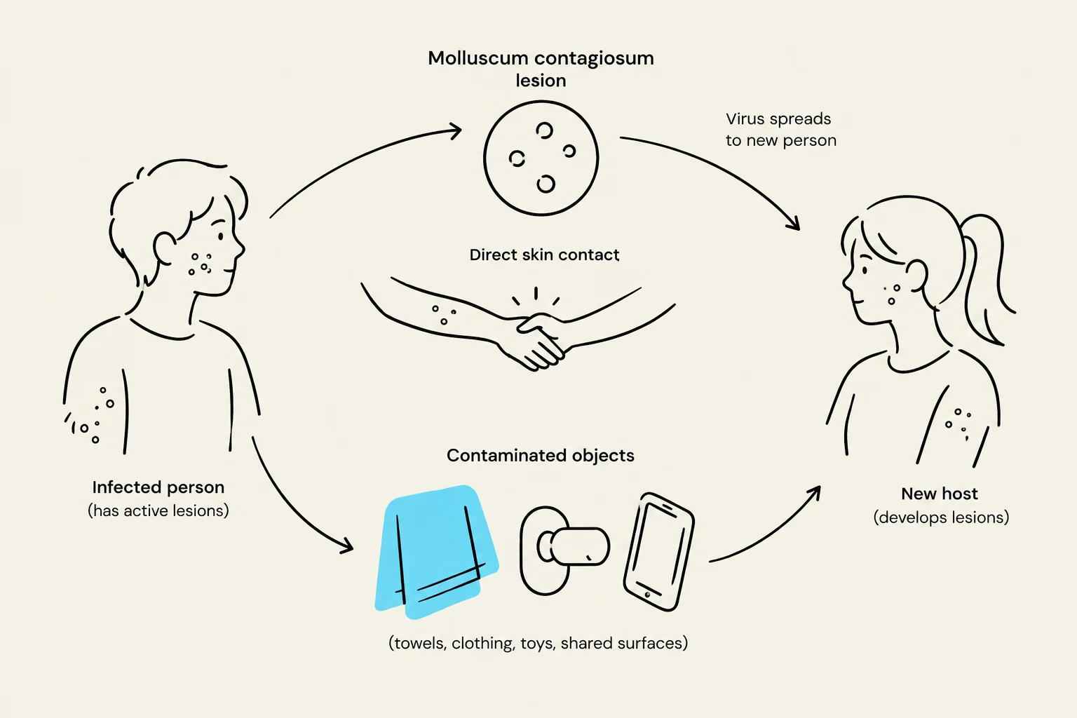
Transmission occurs through direct skin-to-skin contact or via fomites, such as shared towels or gym equipment. In pediatric populations, it is frequently spread through casual contact or shared toys, whereas in adults, sexually transmitted contact is a common route of infection.
Clinical Presentation: The Umbilicated Papule
The most critical skill in diagnosing molluscum contagiosum is the visual identification of its characteristic lesions. The clinical hallmark is the presence of small, firm, dome-shaped papules.
Key Diagnostic Features:
- Umbilication: The most defining feature is a central indentation or "dimple" at the apex of the papule, known as umbilication.
- Appearance: The lesions are typically flesh-colored, pearly, or slightly pink.
- Size and Distribution: They generally range from 2 to 5 mm in diameter. Depending on the mode of transmission, they may appear on the face, trunk, and extremities in children, or in the genital area in adults.

Keep reading for free.
Create an account to finish this reading and make it part of your study routine.
Continue reading freeAlready have an account? Log in
Updated