


Glaucoma is often referred to as the "silent thief of sight" because it frequently progresses without noticeable symptoms until significant, irreversible vision loss has occurred. Understanding the mechanics of the eye, the critical role of intraocular pressure (IOP), and the necessity of strict treatment adherence is essential for anyone studying ocular health or managing the condition. This article explores the physiological relationship between pressure and nerve damage, the challenges of lifelong medication, and the technical aspects of safe administration.
The Relationship Between Intraocular Pressure and Vision Loss
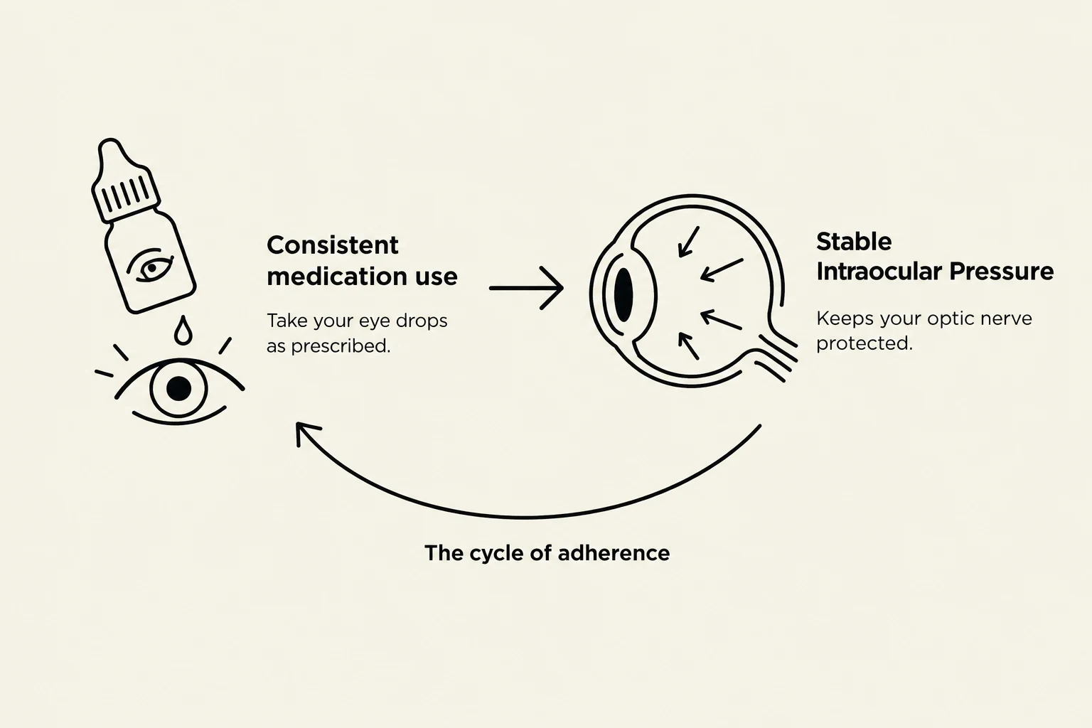
At the core of glaucoma management is the regulation of intraocular pressure (IOP). The eye contains a fluid called aqueous humor, which is continuously produced and drained through a complex network of channels. When the drainage system becomes inefficient, the fluid builds up, leading to an increase in IOP.
This elevated pressure exerts mechanical stress on the optic nerve, which is responsible for transmitting visual information from the retina to the brain. Over time, this stress causes the death of retinal ganglion cells. As these cells die, the optic nerve fibers diminish, leading to progressive peripheral vision loss and, eventually, central blindness.
The relationship between pressure and damage can be conceptualized through the concept of "target pressure." While high pressure is a primary risk factor, the goal of treatment is not necessarily to return pressure to a "normal" range, but to lower it to a level where the optic nerve can remain stable.

Keep reading for free.
Create an account to finish this reading and make it part of your study routine.
Continue reading freeAlready have an account? Log in
Updated