Understanding and Managing Diabetic Emergencies: DKA and HHS
This guide explains the differences between Diabetic Ketoacidosis (DKA) and Hyperosmolar Hyperglycemic Syndrome (HHS), their diagnostic criteria, and essential nursing management strategies, including fluid resuscitation, electrolyte balance, and insulin therapy.
Diabetic emergencies, specifically Diabetic Ketoacidosis (DKA) and Hyperosmolar Hyperglycemic Syndrome (HHS), represent some of the most critical challenges in acute care nursing. While both conditions are driven by extreme hyperglycemia, they arise from different pathophysiological mechanisms and require distinct clinical approaches. Understanding the nuances between the absolute insulin deficiency of DKA and the relative deficiency seen in HHS is vital for preventing fatal complications like cerebral edema or profound electrolyte imbalances.
Diabetic Ketoacidosis (DKA)
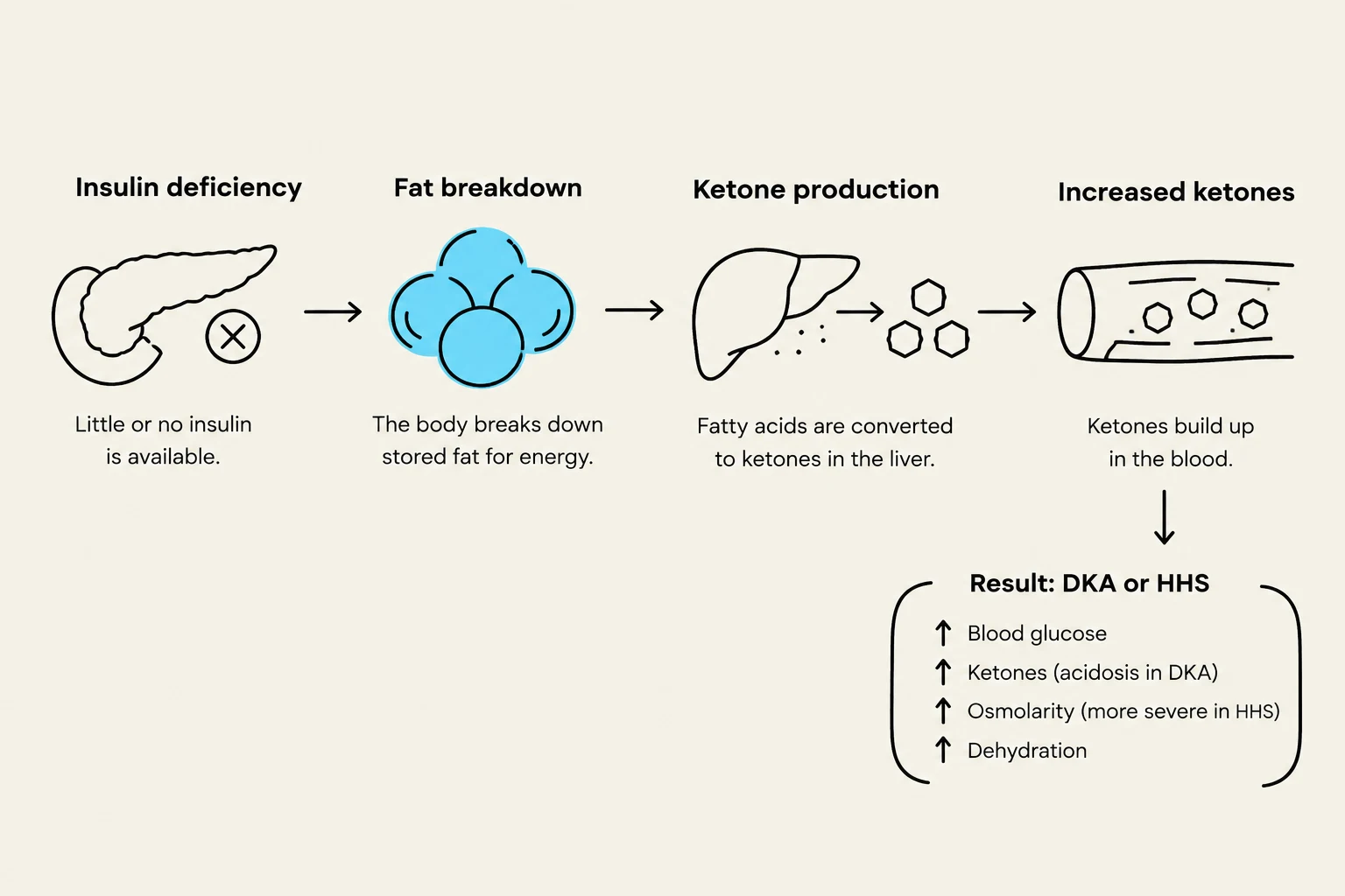
DKA is most commonly associated with Type 1 Diabetes Mellitus, where an absolute lack of insulin forces the body to seek alternative fuel sources. When cells cannot access glucose, the body begins breaking down fats and proteins. This metabolic shift produces acidic byproducts known as ketones.
The clinical presentation of DKA is often dramatic. Patients frequently present with nausea, vomiting, abdominal pain, and polyuria (excessive urination). A hallmark sign is Kussmaul respirations—deep, labored, gasping breaths that serve as a compensatory mechanism to "blow off" CO2 and mitigate metabolic acidosis. Patients may also exhibit "fruity" or acetone-scented breath due to the presence of ketones.
Diagnostic Criteria and the Anion Gap
To confirm a diagnosis of DKA, clinicians look for a specific triad: hyperglycemia, ketosis, and metabolic acidosis. Key laboratory findings include:
A critical diagnostic tool in DKA is the calculation of the anion gap, which measures the concentration of unmeasured anions in the extracellular fluid. An elevated anion gap indicates the presence of metabolic acidosis caused by the accumulation of ketoacids.
Anion Gap=(Na++K+)−(Cl−+HCO3−)
This diagram illustrates the physiological process leading to Diabetic Ketoacidosis (DKA) or Hyperosmolar Hyperglycemic State (HHS) due to insulin deficiency. It shows how insulin deficiency leads to fat breakdown, ketone production in the liver, and increased ketones in the blood, ultimately resulting in elevated blood glucose, ketones, osmolarity, and dehydration.
Hyperosmolar Hyperglycemic Syndrome (HHS)
HHS primarily affects elderly patients with Type 2 Diabetes. Unlike DKA, these patients typically have enough circulating insulin to prevent significant ketone production, but not enough to prevent extreme hyperglycemia.
The hallmark of HHS is extreme hyperglycemia (often >34 mmol/L) and profound dehydration leading to high serum osmolality. Because the hyperglycemia is so severe, patients experience massive osmotic diuresis, leading to severe fluid deficits. This often manifests as neurological changes, including confusion, somnolence, seizures, or even coma.
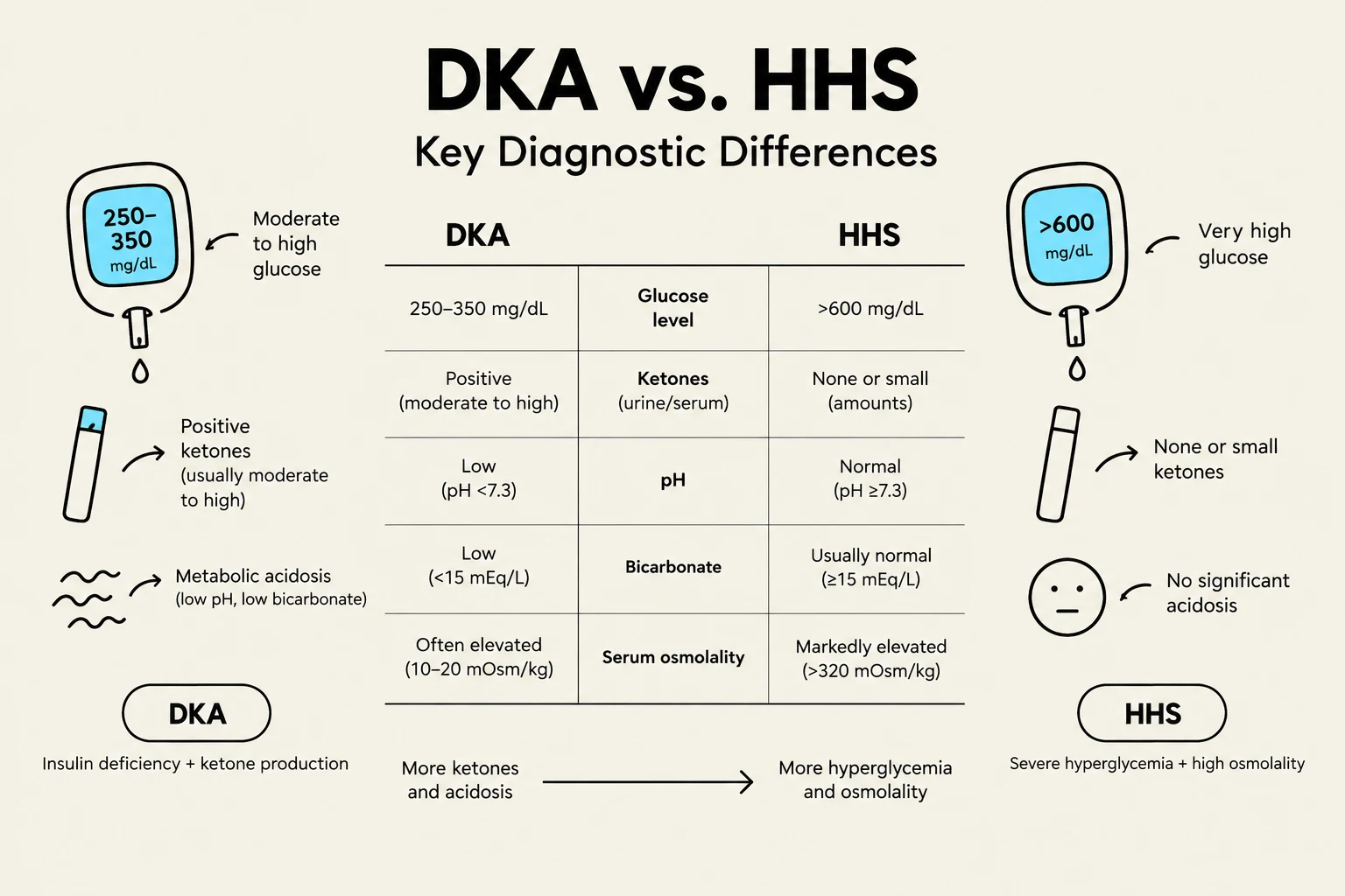
Exam Tip: When comparing the two, remember that HHS is characterized by higher glucose levels and higher osmolality, but minimal to no ketones and no significant acidosis, whereas DKA is defined by the presence of ketones and metabolic acidosis.
Clinical Management: The "D, K, A" Approach
Managing these emergencies requires a systematic approach focusing on Dehydration, Potassium, and Acidosis/Glucose.
1. Dehydration and Fluid Resuscitation
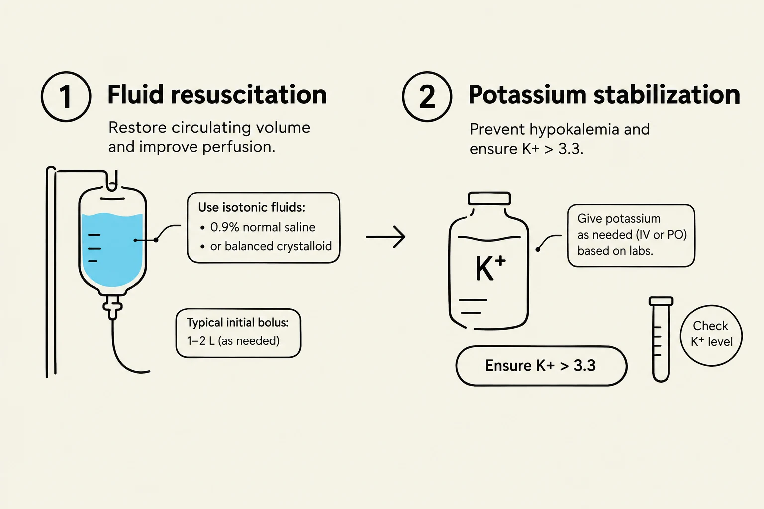
The first priority is restoring circulating volume. In DKA, patients are typically started on isotonic solutions, such as 0.9% Normal Saline (NS).
As blood glucose levels drop, the nurse must monitor for the risk of cerebral edema. To prevent this, once blood glucose reaches approximately 12–14 mmol/L, the provider will typically switch the infusion to a hypotonic solution (e.g., D5 0.5% NS). This prevents a rapid shift of water into brain cells that can occur if glucose is corrected too quickly.
2. Potassium (K+) and Electrolyte Replacement
Insulin therapy causes potassium to shift from the extracellular fluid back into the cells, which can lead to life-threatening hypokalemia.
CRITICAL SAFETY ALERT: In DKA management, insulin must be withheld until the serum potassium (K+) is >3.3 mmol/L. Administering insulin to a patient with low potassium can cause a precipitous drop in serum levels, leading to fatal cardiac dysrhythmias.
Nurses must maintain continuous cardiac monitoring to detect any dysrhythmias resulting from these rapid electrolyte shifts.
3. Acidosis and Insulin Therapy
Once hydration is initiated and potassium levels are safe, insulin therapy is used to suppress ketogenesis and lower blood glucose. Regular insulin is typically administered via continuous IV infusion (e.g., 0.1 unit/kg/hr).
A vital nursing responsibility is ensuring that glucose does not decrease too rapidly. The goal is a gradual reduction—no more than 4 mmol/L per hour. If the drop is too steep, the risk of cerebral edema increases significantly.
This diagram illustrates two steps in medical treatment: fluid resuscitation to restore circulating volume and improve perfusion, and potassium stabilization to prevent hypokalemia. It details the use of isotonic fluids like normal saline or balanced crystalloids for resuscitation, and the administration of potassium based on lab results to maintain levels above 3.3.
Nursing Priorities and Monitoring
For both DKA and HHS, the nurse plays a central role in preventing secondary complications.
Strict Intake and Output (I&O): Patients often require urinary catheterization to monitor hourly output. This is essential to assess renal perfusion and the effectiveness of fluid resuscitation.
Neurological Assessment: Frequent checks of the Level of Consciousness (LOC) using the Glasgow Coma Scale are necessary to detect early signs of cerebral edema or worsening HHS.
Frequent Glucose Monitoring: Blood glucose should be checked frequently (often every 15 minutes to 1 hour) to guide insulin titration and prevent hypoglycemia.
Cardiac Monitoring: Continuous ECG monitoring is mandatory due to the high risk of potassium-induced dysrhythmias.
Feature
Diabetic Ketoacidosis (DKA)
Hyperosmolar Hyperglycemic Syndrome (HHS)
Primary Population
Type 1 Diabetes
Type 2 Diabetes (often elderly)
Blood Glucose
>15 mmol/L
>34 mmol/L
Ketones
Positive (Serum/Urine)
Absent or minimal
Acidosis
Present (pH <7.3)
Absent
Dehydration
Moderate to Severe
Extreme (often >10L deficit)
This chart compares the key diagnostic differences between Diabetic Ketoacidosis (DKA) and Hyperosmolar Hyperglycemic State (HHS), focusing on glucose levels, ketones, pH, bicarbonate, and serum osmolality.
Keep reading for free.
Create an account to finish this reading and make it part of your study routine.