Understanding and Managing Diabetes Mellitus Types 1 and 2
This content provides a comprehensive overview of Diabetes Mellitus, differentiating between Type 1 and Type 2. It covers pathophysiology, diagnostic criteria, clinical manifestations like hypoglycemia and hyperglycemia, and management strategies including lifestyle modifications, oral medications, and insulin therapy.
Diabetes mellitus is a group of metabolic disorders characterized by chronic hyperglycemia resulting from defects in insulin secretion, insulin action, or both. Understanding the distinction between Type 1 and Type 2 diabetes is fundamental to clinical management, as the underlying pathophysiology dictates whether a patient requires lifelong insulin therapy or can be managed through lifestyle modifications and oral medications.
Pathophysiology: Type 1 vs. Type 2 Diabetes
The primary difference between the two types lies in the mechanism of insulin deficiency.
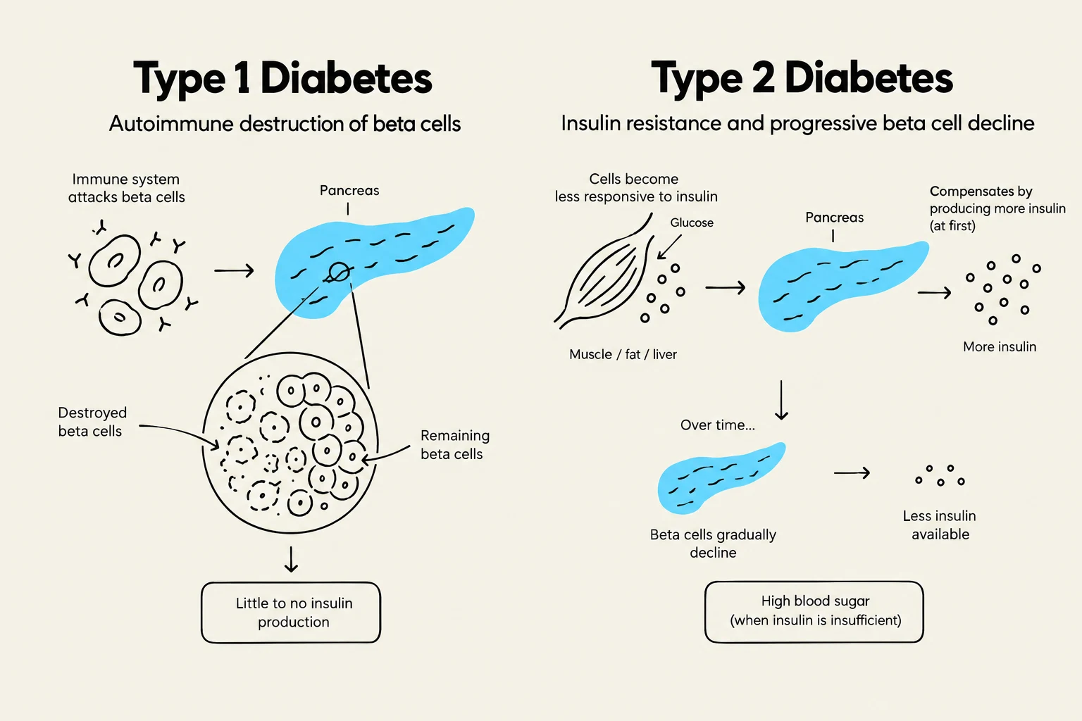
Type 1 Diabetes Mellitus is an autoimmune condition where the body's immune system destroys the insulin-producing beta cells in the pancreas. This results in an absolute insulin deficiency, meaning the patient cannot produce any insulin and requires exogenous insulin for survival. It typically has an earlier onset in life.
Type 2 Diabetes Mellitus is characterized by insulin resistance—where cells do not respond effectively to insulin—and hyperinsulinemia in the early stages. Over time, the pancreas may fail to produce enough insulin to overcome this resistance, leading to a relative insulin deficiency. While traditionally associated with adult onset, it is increasingly being diagnosed in younger populations.
Diagram illustrating the differences between Type 1 and Type 2 Diabetes, focusing on beta cell function and insulin production.
Diagnostic Criteria and Glycemic Targets
Clinicians rely on specific biochemical thresholds to diagnose diabetes and identify pre-diabetes.
The HbA1C (glycated hemoglobin) test is particularly valuable because it reflects the mean plasma glucose level over the previous 90–120 days, corresponding to the lifespan of a red blood cell.
Individualized Glycemic Control
Once diagnosed, the goal is to maintain glycemic control to prevent long-term complications. While a general target for HbA1C is ≤7.0%, targets must be individualized based on the patient's clinical status:
Non-critically ill patients: Aim for $5–10$ mmol/L post-meal.
Critically ill patients: Aim for $8–10$ mmol/L.
Frail elderly patients: May have higher targets of $5–12$ mmol/L to avoid the dangers of hypoglycemia.
Clinical Manifestations: Hypoglycemia and Hyperglycemia
Blood glucose fluctuations can lead to acute, life-threatening emergencies.
Hypoglycemia (Low Blood Sugar)
Defined as blood glucose <4 mmol/L, hypoglycemia can be caused by excessive insulin, insufficient food intake, or inconsistent exercise.
Symptoms: Shaking, sweating, palpitations, anxiety, hunger, confusion, and dizziness.
Severe cases: May progress to seizures, coma, or death.
Nursing Alert: The 15/15 Rule
For a conscious patient, treat hypoglycemia with 15g of fast-acting carbohydrates (e.g., 4 glucose tablets or 125ml of apple juice). Re-check blood glucose after 15 minutes. If still <4 mmol/L, repeat the 15g dose.
Hyperglycemia (High Blood Sugar)
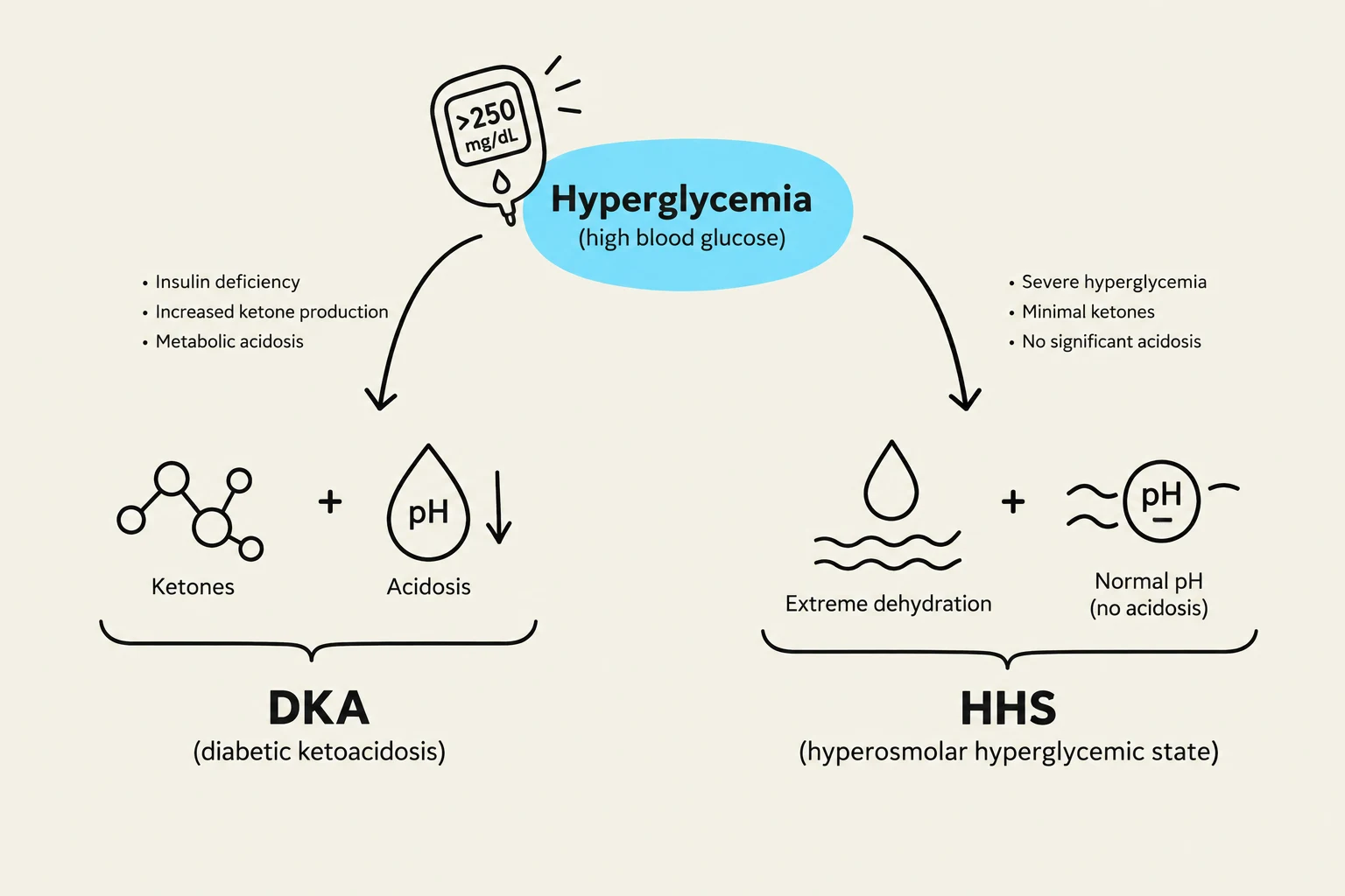
Symptoms of hyperglycemia (>7 mmol/L) include polyphagia (increased hunger), polydipsia (increased thirst), polyuria (increased urination), blurred vision, and lethargy. If left untreated, this can progress to two major emergencies: Diabetic Ketoacidosis (DKA) or Hyperglycemic Hyperosmolar State (HHS).
This diagram illustrates the differences between Diabetic Ketoacidosis (DKA) and Hyperosmolar Hyperglycemic State (HHS), both conditions associated with hyperglycemia (high blood glucose).
Management of Type 2 Diabetes
Management of Type 2 diabetes focuses on lifestyle changes and pharmacological intervention to improve insulin sensitivity and reduce glucose production.
Nutritional Therapy: A diet high in fiber, lean proteins, and healthy fats (like monounsaturated fats) is recommended. Patients should prioritize low-glycemic foods to delay glucose release into the bloodstream.
Physical Activity: Aim for at least 150 minutes of moderate aerobic activity per week, supplemented by resistance training 2–3 times per week.
Oral Hypoglycemic Agents (OHA):
Biguanides (e.g., Metformin): Decreases hepatic glucose production and increases insulin sensitivity.
GLP-1 Analogues (e.g., Ozempic/Semaglutide): Suppresses appetite and regulates insulin secretion.
SGLT2 Inhibitors: Promote glucose excretion via the urine.
Insulin Therapy and Hospital Management
Type 1 diabetics require exogenous insulin, and some Type 2 diabetics may require insulin if oral medications are insufficient. In a hospital setting, Basal Bolus Insulin Therapy (BBIT) is often utilized to mimic physiological insulin patterns.
The BBIT Framework
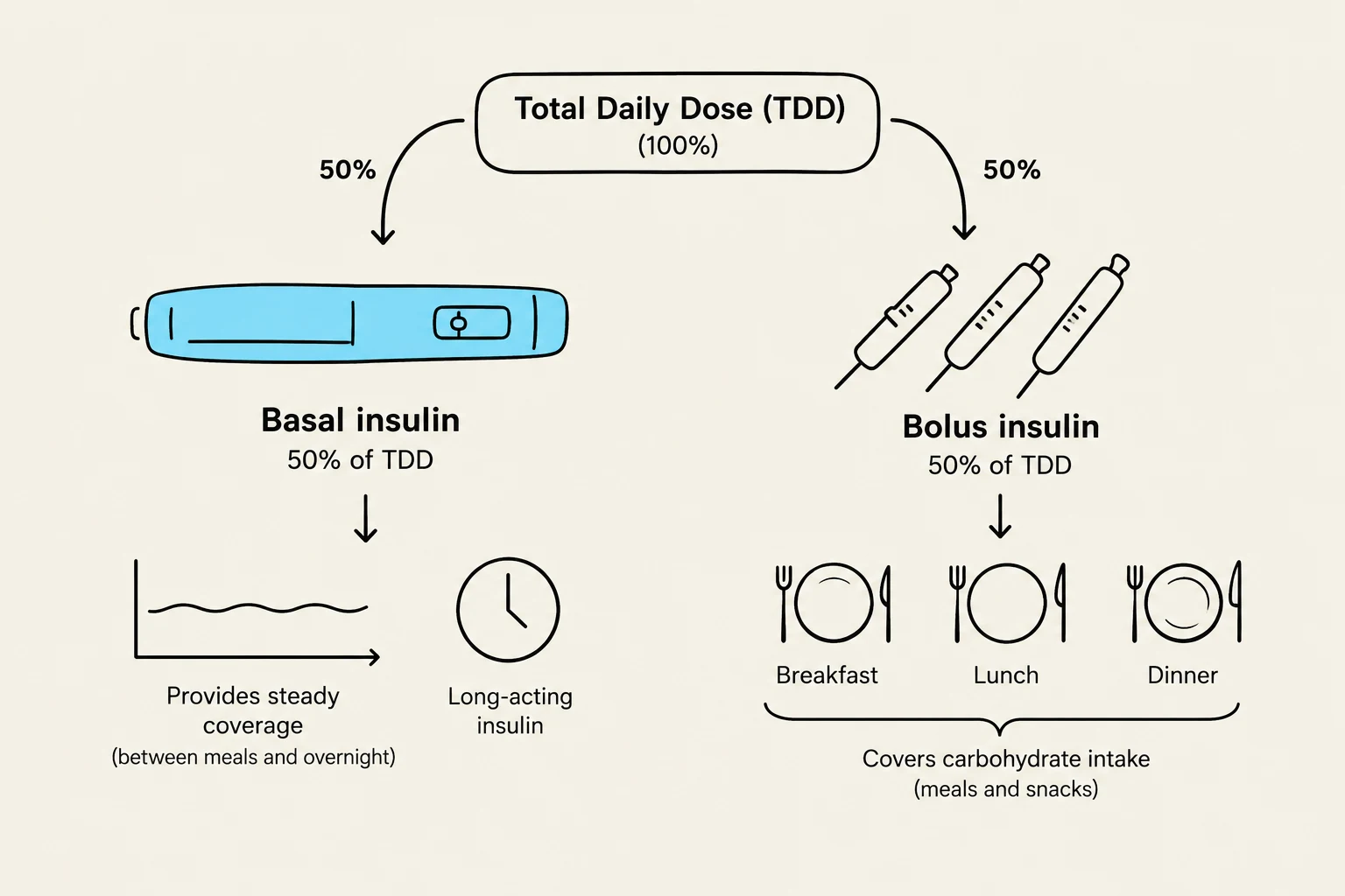
The Total Daily Dose (TDD) is typically split into two main components:
Basal Insulin (50% of TDD): Long-acting insulin (e.g., Lantus) that covers overnight and between-meal glucose levels. It is given even if the patient is NPO (nothing by mouth) to prevent DKA.
Bolus Insulin (50% of TDD): Rapid-acting insulin (e.g., Aspart or Lispro) divided among meals to cover post-prandial glucose spikes. Note: Bolus insulin must be held if the patient is NPO.
Correction Insulin (Sliding Scale): Additional insulin administered at mealtimes to correct hyperglycemia based on the current blood glucose reading.
This diagram illustrates the breakdown of Total Daily Dose (TDD) insulin into basal and bolus insulin. Basal insulin, comprising 50% of the TDD, provides steady coverage between meals and overnight, often administered as long-acting insulin. Bolus insulin, also 50% of the TDD, is used to cover carbohydrate intake during meals and snacks, typically administered before breakfast, lunch, and dinner.
Complications of Therapy and Acute Emergencies
Insulin-Related Complications
Somogyi Effect: Rebound morning hyperglycemia caused by undetected hypoglycemia during the night.
Dawn Phenomenon: Morning hyperglycemia caused by natural surges in counter-regulatory hormones like cortisol.
Lipodystrophy: Tissue changes (hypertrophy or atrophy) at injection sites that impair insulin absorption.
DKA vs. HHS
When blood glucose levels become extremely high, patients may enter a critical state.
Feature
Diabetic Ketoacidosis (DKA)
Hyperglycemic Hyperosmolar State (HHS)
Primary Type
Type 1
Type 2 (often elderly)
Glucose Level
High (>15 mmol/L)
Extremely High (>34 mmol/L)
Ketones
Present (Positive urine/serum)
Absent or minimal
Acidosis
Metabolic Acidosis (pH <7.3)
Absent (Normal Bicarbonate)
Key Risk
Dehydration, electrolyte shift
Severe dehydration, neurological deficit
Critical Management Rule for DKA: When treating DKA with insulin, clinicians must ensure the patient's potassium ($K^+$) level is >3.3 mmol/L before starting insulin infusion, as insulin will drive potassium into the cells and can cause profound hypokalemia. Furthermore, glucose must be corrected slowly (no more than 4 mmol/L per hour) to prevent cerebral edema.
Keep reading for free.
Create an account to finish this reading and make it part of your study routine.