A comprehensive overview of Type 1 and Type 2 diabetes mellitus, covering pathophysiology, diagnostic criteria, glycemic targets, insulin therapy, complications, and lifestyle management.
Diabetes mellitus is a complex metabolic disorder characterized by chronic hyperglycemia resulting from defects in insulin secretion, insulin action, or both. Effective management requires a deep understanding of the underlying pathophysiology, precise diagnostic monitoring, and highly individualized therapeutic strategies to prevent life-threatening acute complications.
Pathophysiology: Type 1 vs. Type 2 Diabetes
While both types result in elevated blood glucose, their origins are fundamentally different.
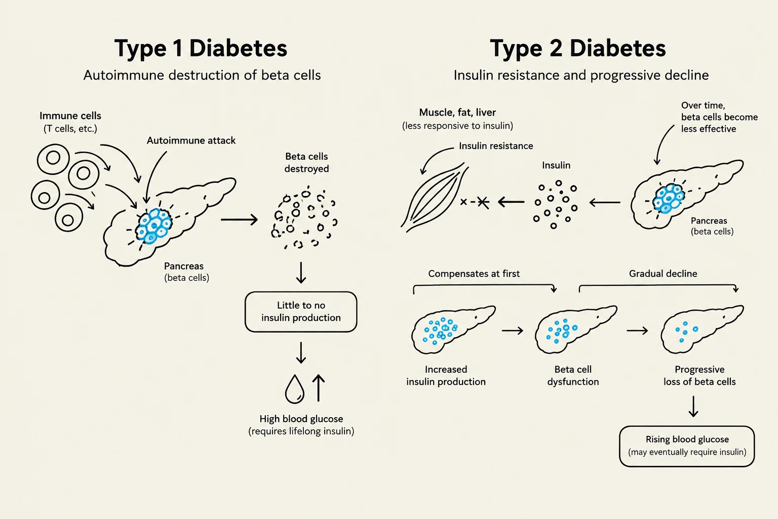
Type 1 Diabetes Mellitus is characterized by the autoimmune destruction of pancreatic beta cells. This leads to an absolute insulin deficiency, meaning the body produces little to no insulin. Consequently, patients with Type 1 diabetes require exogenous insulin for survival.
Type 2 Diabetes Mellitus typically involves a progression from insulin resistance (where cells do not respond effectively to insulin) to hyperinsulinemia, and eventually to a relative decrease in insulin production. While often associated with adult onset, it is increasingly seen in younger populations. Management often begins with lifestyle modifications and oral medications, though insulin may be required in later stages.
This illustration compares Type 1 and Type 2 diabetes, detailing the autoimmune destruction of beta cells in Type 1 and insulin resistance leading to beta cell decline in Type 2.
Diagnostic Criteria and Glycemic Targets
To diagnose diabetes, clinicians rely on several key metrics: Fasting Glucose (FG), HbA1C (glycated hemoglobin), and the Oral Glucose Tolerance Test (OGTT).
Diagnostic Thresholds
Metric
Normal
Prediabetes
Diabetes
Fasting Glucose (FG)
<5.6 mmol/L
6.1−6.9 mmol/L
≥7.0 mmol/L
HbA1C
<6.0%
$6.0 - 6.4%$
>6.5%
2-hr OGTT
5.0−10.0 mmol/L
7.8−11.0 mmol/L
≥11.1 mmol/L
Random Glucose
N/A
N/A
≥11.1 mmol/L
Note: HbA1C reflects the mean plasma glucose level over the previous 90–120 days, corresponding to the lifespan of a red blood cell.
Individualized Glycemic Targets
A general target for most patients is an HbA1C≤7.0%. However, targets must be individualized based on the patient's clinical status:
Non-critical illness:5–10 mmol/L.
Critical illness:8–10 mmol/L.
Frail elderly:5–12 mmol/L.
Clinical Manifestations: Hypoglycemia and Hyperglycemia
Patients must be able to recognize the symptoms of glucose instability to prevent emergencies.
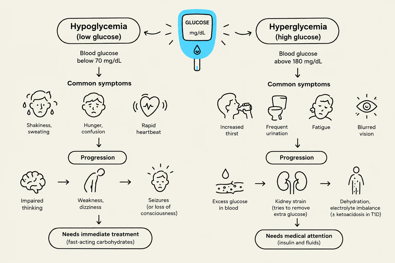
Hypoglycemia (<4 mmol/L)
Hypoglycemia (low blood sugar) can be caused by excessive insulin, insufficient food intake, or inconsistent exercise. Symptoms include shaking, sweating, palpitations, anxiety, hunger, confusion, and dizziness. If untreated, it can progress to seizures, coma, or death.
Hyperglycemia (>7 mmol/L)
Hyperglycemia (high blood sugar) presents with the "three Ps": Polyphagia (hunger), polydipsia (thirst), and polyuria (excessive urination). Other signs include blurred vision, dry skin, and lethargy. Severe hyperglycemia can lead to life-threatening states like Diabetic Ketoacidosis (DKA) or Hyperglycemic Hyperosmolar State (HHS).
This diagram illustrates the symptoms and progression of hypoglycemia (low blood glucose) and hyperglycemia (high blood glucose), along with their respective treatments.
Insulin Therapy and Management
For many patients, particularly those with Type 1 or advanced Type 2, Basal Bolus Insulin Therapy (BBIT) is the gold standard for mimicking physiological insulin release.
The Components of BBIT
Basal Insulin: Long-acting (e.g., Glargine) or intermediate-acting (e.g., NPH) insulin. It stabilizes glucose overnight and between meals by covering the glucose produced by the liver.
Bolus Insulin: Rapid-acting (e.g., Aspart, Lispro) insulin administered at mealtimes to cover glucose spikes from carbohydrate ingestion.
Correctional Insulin: Often administered via a "sliding scale" at mealtimes to address pre-meal glucose levels that are higher than the target.
Calculating the Total Daily Dose (TDD)
In a clinical setting, a physician determines the TDD based on the patient's weight and insulin sensitivity:
Type 1 or Type 2 with renal dysfunction:TDD=Weight (kg)⋅(0.3 to 0.5 units/kg/day)
New-start Type 2 (insulin resistance):TDD=Weight (kg)⋅(0.5 to 1.0 units/kg/day)
Once the TDD is established, it is typically split: approximately 50% for Basal insulin and 50% for Bolus insulin.
Exam Tip: Always check blood glucose levels before administering bolus insulin. If a patient is NPO (nothing by mouth), bolus insulin should be held to prevent severe hypoglycemia.
Complications of Therapy and Disease
Insulin-Related Complications
Somogyi Effect: Undetected nocturnal hypoglycemia followed by rebound morning hyperglycemia. This is often treated by reducing the evening insulin dose.
Dawn Phenomenon: Morning hyperglycemia caused by the natural surge of counter-regulatory hormones (growth hormone, cortisol). This is treated by adjusting the timing of insulin or increasing the dose.
Lipodystrophy: Tissue changes (hypertrophy or atrophy) at injection sites due to repeated use of the same area, which can impair insulin absorption.
Acute Metabolic Emergencies
Feature
Diabetic Ketoacidosis (DKA)
Hyperglycemic Hyperosmolar State (HHS)
Primary Type
Type 1 (Absolute deficiency)
Type 2 (Relative deficiency)
Glucose Level
High (>15 mmol/L)
Very High (>34 mmol/L)
Ketones
Present in blood/urine
Absent or minimal
Acidosis
Present (pH <7.3)
Absent (Normal pH/Bicarb)
Key Risk
Metabolic Acidosis
Severe Dehydration/Osmolality
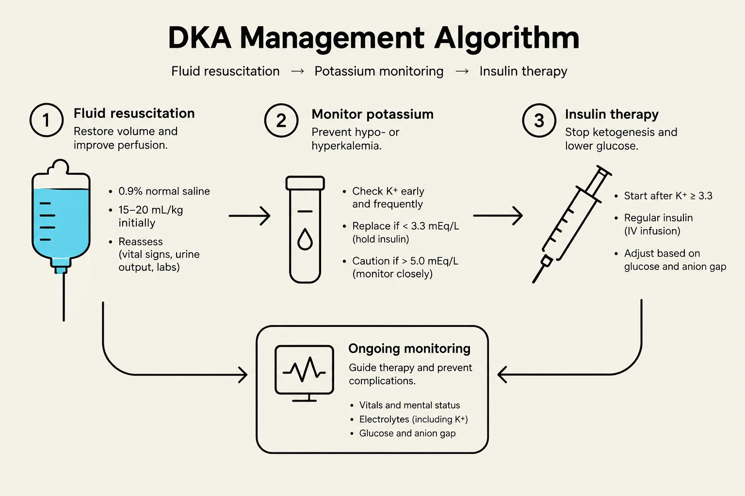
Management of DKA requires a careful balance of fluids, potassium, and insulin. A critical rule is that insulin should not be started until the serum potassium ($K^+$) is ≥3.3 mmol/L to prevent life-threatening hypokalemia. Furthermore, glucose must be reduced gradually (no more than 4 mmol/L per hour) to prevent cerebral edema.
Management of HHS focuses heavily on massive fluid resuscitation, as patients often have a deficit of over 10L of fluid.
This algorithm outlines the management of Diabetic Ketoacidosis (DKA), detailing steps for fluid resuscitation, potassium monitoring, and insulin therapy, along with ongoing monitoring of vital signs, electrolytes, and glucose.
Lifestyle and Self-Management
For Type 2 diabetes, management is anchored in lifestyle changes:
Nutrition: High fiber to delay glucose release, lean proteins, and healthy fats (MUFAs/PUFAs).
Physical Activity: At least 150 minutes of moderate activity per week, such as brisk walking.
Monitoring: Regular blood glucose testing and, when ill, increased frequency (every 2–4 hours) and urine ketone testing to prevent DKA.
Keep reading for free.
Create an account to finish this reading and make it part of your study routine.