This guide explains the pathophysiology, clinical phases, and nursing interventions for Acute Kidney Injury (AKI), Chronic Kidney Disease (CKD), and Polycystic Kidney Disease (PKD). It also covers renal replacement therapies like hemodialysis and peritoneal dialysis.
The kidneys are vital organs responsible for maintaining homeostasis through the excretion of waste, regulation of fluid and electrolyte balance, acid-base regulation, blood pressure control, and the secretion of erythropoietin for red blood cell production. When these functions are compromised, patients may face life-threatening complications. Understanding the distinctions between acute and chronic renal dysfunction, as well as specific genetic conditions like Polycystic Kidney Disease, is essential for effective nursing management.
Acute Kidney Injury (AKI)
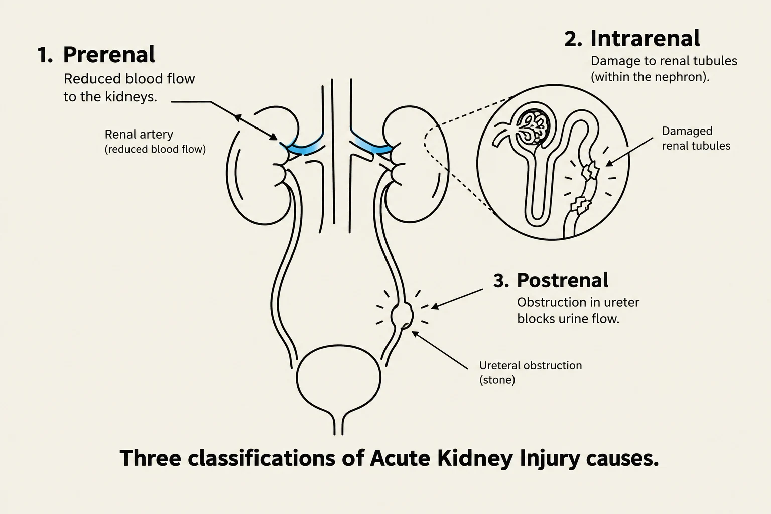
This diagram illustrates the three main classifications of acute kidney injury: prerenal (reduced blood flow to the kidneys), intrarenal (damage to renal tubules within the nephron), and postrenal (obstruction in the ureter blocking urine flow).
Acute Kidney Injury (AKI), formerly known as acute renal failure, is characterized by a rapid and progressive loss of renal function. This condition often leads to azotemia, which is the accumulation of nitrogenous waste products in the blood.
Pathophysiology and Causes
AKI is generally categorized into three major causes based on the location of the insult:
Prerenal: The most common cause, resulting from external factors that reduce renal blood flow (e.g., hypovolemia, decreased cardiac output, sepsis, or hemorrhage).
Intrarenal: Direct damage to the renal parenchymal tissues, such as the nephrons. Acute Tubular Necrosis (ATN) accounts for 90% of all intrarenal cases.
Postrenal: Mechanical obstruction of the lower urinary tract, such as in the ureters, bladder, or urethra.
Clinical Phases of AKI
AKI typically progresses through four distinct phases:
Initiating (Onset) Phase: Lasts hours to days; involves a decrease in circulating volume and compensatory vasoconstriction.
Oliguric Phase: Characterized by oliguria, or urine output below 400 mL/day. Patients may experience fluid overload, metabolic acidosis, and electrolyte imbalances.
Diuretic Phase: As the cause is corrected, urine output increases significantly (1–3 L or even 3–5 L per day) due to osmotic diuresis from high urea levels. This phase carries a risk of hypovolemia and hypotension.
Recovery Phase: The kidneys return to regular function, and the Glomerular Filtration Rate (GFR) increases to 70–80% of normal.
Tip of the Day: In the oliguric phase of AKI, urine output is <400 mL/day. In the diuretic phase, output can reach as high as 5 L/day.
Nursing Interventions for AKI
Nursing care focuses on eliminating the cause and preventing complications. Key actions include:
Fluid Management: Monitoring strict Intake and Output (I&O) and daily weights.
Electrolyte Monitoring: Watching for life-threatening hyperkalemia (high potassium), which can cause cardiac dysrhythmias such as peaked T waves or even asystole.
Pharmacology: Administering diuretics (e.g., Furosemide) to promote filtration, or potassium-lowering therapies like Kayexalate or IV insulin/glucose in emergencies.
Chronic Kidney Disease (CKD)
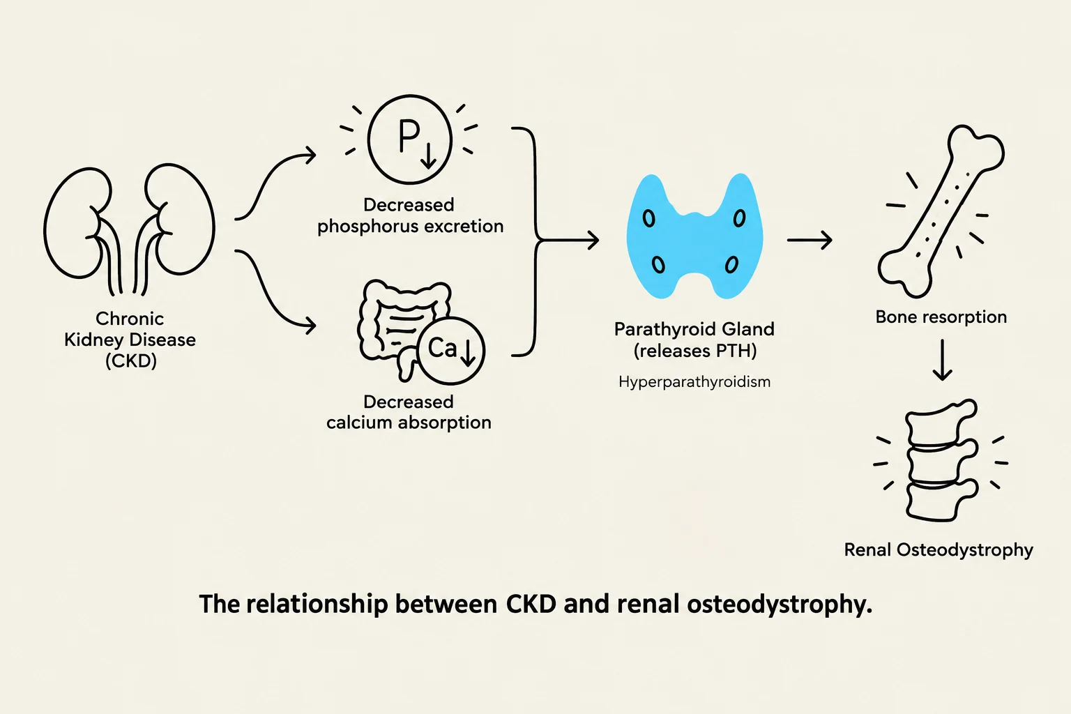
This diagram illustrates the relationship between Chronic Kidney Disease (CKD) and Renal Osteodystrophy, showing how CKD leads to decreased phosphorus excretion and calcium absorption, which in turn affects the parathyroid gland and results in bone resorption.
Unlike the rapid onset of AKI, Chronic Kidney Disease (CKD) is a progressive, irreversible, and slow decline in renal function. It is often caused by long-term comorbidities such as uncontrolled diabetes and hypertension.
Diagnostic Indicators
The severity of CKD is staged based on the Glomerular Filtration Rate (GFR) and the presence of protein in the urine. According to the National Kidney Foundation:
Normal: eGFR ≥90.
Early-stage disease: eGFR 60–89.
Kidney disease: eGFR 15–59.
Kidney failure: eGFR <15.
Clinical Manifestations and Complications
CKD patients often present with anemia (due to decreased erythropoietin production), hypertension, and fluid retention.
A significant complication is Renal Osteodystrophy, which results from the inability to maintain calcium and phosphorus balance. This leads to hyperparathyroidism and hypocalcemia. Patients may exhibit neuromuscular irritability, such as Chvostek's or Trousseau's signs.
Management of CKD
Nutrition: Essential to slow progression. This includes protein restriction (in stages 1–4), phosphorus restriction, and sodium/potassium limits.
Medication Safety: Because excretion is delayed, dosages of medications like antibiotics or NSAIDs must be adjusted to avoid toxicity.
Renal Replacement Therapies (RRTs): When the kidneys can no longer sustain life, RRTs are required.
Polycystic Kidney Disease (PKD)
Polycystic Kidney Disease is a congenital, genetic disorder characterized by the development of multiple fluid-filled cysts within the nephrons. As these cysts grow, they replace healthy kidney tissue, leading to progressive renal failure.
Clinical Features
PKD can manifest in childhood or as an adult-onset (ADPKD) condition. Common clinical manifestations include:
Hypertension.
Hematuria (blood in urine).
Back or flank pain.
Enlarged kidneys and increased abdominal girth.
Approximately 50% of the population with PKD progresses to End-Stage Renal Disease (ESRD) by age 60.
Nursing Care for PKD
Management focuses on symptom control and preventing complications:
Pain Management: Addressing flank and back pain.
Infection Prevention: Monitoring for signs of UTI.
Therapeutic Procedures: In some cases, needle aspiration may be required to drain large cysts.
Renal Replacement Therapies (RRTs)
This diagram illustrates the mechanisms of hemodialysis and peritoneal dialysis, comparing the two methods of kidney dialysis.
When renal function declines to ESRD, patients require RRTs to perform the work of the kidneys.
Hemodialysis: A process where blood is filtered through an external machine. Access is often created via an Arteriovenous (AV) Fistula or graft.
Peritoneal Dialysis (PD): Uses the patient's peritoneal membrane as a filter by instilling dialysis fluid into the abdominal cavity.
Summary Table: Laboratory Values for Renal Function
Test
Normal Range
Significance
Blood Urea Nitrogen (BUN)
8–21 mg/dL
Measures renal excretion of urea nitrogen
Serum Creatinine
0.5–1.2 mg/dL
Primary indicator of kidney function
Keep reading for free.
Create an account to finish this reading and make it part of your study routine.