Understanding Acid-Base Balance and Arterial Blood Gas Interpretation
This guide explains the physiological mechanisms of homeostasis, focusing on acid-base balance regulated by buffer, respiratory, and renal systems. It details the components of Arterial Blood Gas (ABG) analysis, identifies common acid-base imbalances (respiratory and metabolic acidosis/alkalosis), and outlines a step-b
Maintaining a stable internal environment is the cornerstone of human physiology. This state of equilibrium, known as homeostasis, ensures that despite external changes, the body's internal conditions—such as fluid volume, electrolyte concentrations, and pH—remain within a narrow, functional range. Among these, acid-base balance is perhaps the most critical for cellular function; if the body becomes too acidic or too alkaline, vital enzymatic and metabolic processes can fail.
The Mechanisms of Homeostasis
The body manages pH through a sophisticated hierarchy of responses involving three primary systems:
The Buffer Systems: These act as the first line of defense, providing an immediate chemical response to neutralize acids or bases.
The Respiratory System: This system provides a fast response (within minutes) by adjusting the excretion of carbon dioxide (CO2), which acts as a respiratory acid.
The Renal System: The kidneys provide the most powerful, albeit slowest, response (taking hours to days). They regulate pH by excreting hydrogen ions ($H^+$) and reabsorbing bicarbonate (HCO3−).
Additionally, the Endocrine System supports fluid and electrolyte balance—which are intrinsically linked to acid-base stability—by releasing hormones such as aldosterone and antidiuretic hormone (ADH).
Primary Buffer Systems
To resist sudden changes in pH, the body utilizes several chemical buffers:
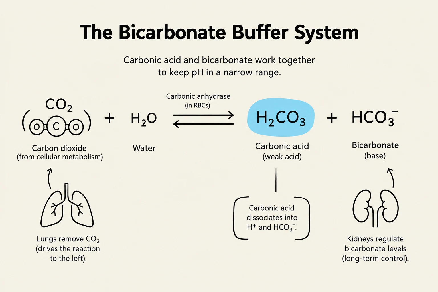
Bicarbonate Buffer System: The most important extracellular buffer, balancing carbonic acid (H2CO3) and bicarbonate (HCO3−).
Protein Buffers: Includes hemoglobin in red blood cells and various plasma proteins that can bind to $H^+$ ions.
Phosphate Buffers: Primarily active within the renal system to regulate intracellular and tubular pH.
The bicarbonate buffer system illustrates how carbonic acid and bicarbonate maintain pH balance. Carbon dioxide from cellular metabolism reacts with water, catalyzed by carbonic anhydrase, to form carbonic acid. This acid can dissociate into H+ and bicarbonate. The lungs remove CO2, influencing the reaction, while the kidneys regulate bicarbonate levels for long-term control.
Understanding Arterial Blood Gas (ABG) Components
To assess a patient's acid-base status, clinicians analyze Arterial Blood Gas (ABG) values. There are four key components to monitor:
Component
Normal Range
Primary Role
pH
7.35 – 7.45
Measures acidity vs. alkalinity
PaCO2
35 – 45 mmHg
The respiratory component (acid)
HCO3−
22 – 26 mEq/L
The metabolic component (base)
PaO2
80 – 100 mmHg
Measures oxygenation
Exam Tip: Remember that pH is the "master" value. If the pH is outside 7.35–7.45, the patient is in a state of imbalance (acidosis or alkalosis).
Identifying Acid-Base Imbalances
An imbalance is classified as either Acidosis (low pH) or Alkalosis (high pH). The root cause determines whether the imbalance is respiratory or metabolic.
Respiratory Imbalances
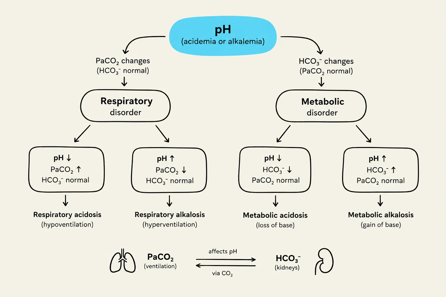
These are caused by changes in ventilation and CO2 levels. In respiratory issues, the pH and PaCO2 move in opposite directions:
Respiratory Acidosis: Low pH (< 7.35) and high PaCO2 (> 45 mmHg). This is typically caused by hypoventilation (retaining too much CO2).
Respiratory Alkalosis: High pH (> 7.45) and low PaCO2 (< 35 mmHg). This is typically caused by hyperventilation (blowing off too much CO2).
Metabolic Imbalances
These are caused by changes in bicarbonate levels. In metabolic issues, the pH and HCO3− move in the same direction:
Metabolic Alkalosis: High pH (> 7.45) and high HCO3− (> 26 mEq/L).
This diagram illustrates the classification of acid-base disorders, differentiating between respiratory and metabolic causes based on changes in pH, PaCO2, and HCO3 levels.
Determining Compensation Status
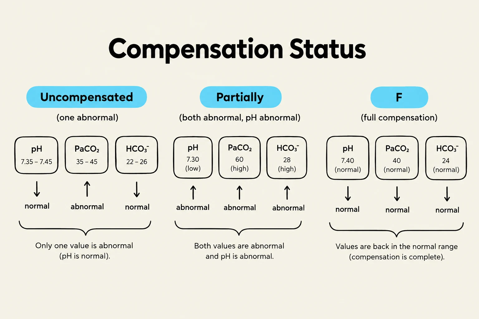
When one system (e.g., the lungs) fails, the other system (e.g., the kidneys) attempts to compensate to bring the pH back toward normal. We categorize this into three stages:
Uncompensated: The pH is abnormal, and only one component (PaCO2 or HCO3−) is abnormal. The other system is still within the normal range, meaning it has not yet begun to help.
Partially Compensated: The pH is still abnormal, but both the PaCO2 and HCO3− are abnormal. This indicates that the secondary system is actively working to correct the pH.
Fully Compensated: The pH has returned to the normal range (7.35–7.45), but both PaCO2 and HCO3− remain abnormal.
Step-by-Step Interpretation Guide
To interpret an ABG, follow this 4-step approach:
Look at pH: Is it Acidosis (< 7.35) or Alkalosis (> 7.45)?
Check PaCO2: Does this match the pH (Respiratory)?
Check HCO3−: Does this match the pH (Metabolic)?
Determine Compensation: Is the other system moving to help?
Worked Examples
Let's apply the rules to these practice scenarios:
Example 1: pH 7.30, PaCO2 50, HCO3− 24
pH is low → Acidosis.
PaCO2 is high (50) → This matches the acidosis (Respiratory).
HCO3− is normal (24) → No compensation yet.
Result: Uncompensated Respiratory Acidosis.
Example 2: pH 7.37, PaCO2 55, HCO3− 31
pH is normal (but on the acidic side of 7.40) → Fully Compensated.
PaCO2 is high (55) → Respiratory component.
HCO3− is high (31) → Metabolic component is compensating.
Result: Fully Compensated Respiratory Acidosis.
This diagram illustrates the three states of compensation status in relation to pH, PaCO2, and HCO3 levels: uncompensated, partially compensated, and fully compensated.
Final Summary Rule:
Respiratory: pH and CO2 move in opposite directions.
Metabolic: pH and HCO3− move in the same direction.
Keep reading for free.
Create an account to finish this reading and make it part of your study routine.