Understanding A and V Pattern Strabismus: Causes and Clinical Presentation
This document explains A and V patterns in strabismus, detailing their definitions, causes related to muscle function and anatomy, and associated syndromes. It covers the distinction between pathological and physiological patterns and provides a summary table of aetiologies.
In clinical strabismus, the magnitude of horizontal deviation often changes depending on the direction of gaze. When these changes are significant, they are classified as "alphabet patterns." Understanding the underlying aetiology of these patterns is critical, as the cause—whether it stems from oblique muscle over-action, vertical muscle weakness, or anatomical insertion anomalies—directly dictates the surgical management strategy.
This graph compares the horizontal deviation in prism diopters for V and A patterns across different gaze directions, including depression, primary, and elevation. It illustrates increased convergence in elevation for the A pattern and increased divergence in elevation for the V pattern.
Defining Alphabet Patterns
Correlation between palpebral fissure slant and pattern type, illustrating downward-slanting (antimongoloid) associated with V patterns and upward-slanting (mongoloid) associated with A patterns.
Alphabet patterns are categorized based on the primary horizontal deviation (esotropia or exotropia) and the shape of the deviation curve.
V Patterns: Characterized by increased divergence on elevation compared to depression. A V-esotropia (V-eso) shows more convergence in depression, while a V-exotropia (V-exo) shows more divergence in elevation.
A Patterns: Characterized by increased convergence on elevation compared to depression. An A-esotropia (A-eso) shows more convergence in elevation, while an A-exotropia (A-exo) shows more divergence in depression.
Pathological vs. Physiological
It is important to distinguish between a normal physiological tendency and a pathological pattern:
Physiological V pattern: A tendency for divergence on elevation where the difference between depression and elevation is <15Δ.
Pathological V pattern: A difference >15Δ.
Pathological A pattern: A difference between elevation and depression >10Δ.
Clinical Pearl: Palpebral Fissure Slant
There is a notable correlation between eyelid morphology and pattern type. Downward slanting (antimongoloid) palpebral fissures are frequently associated with V patterns, whereas upward slanting (mongoloid) fissures are associated with A patterns.
Aetiology: Why do patterns occur?
The aetiology of pattern strabismus is multifactorial. While there is disagreement in the literature regarding the single most common cause, several major theories exist.
1. Abnormal Oblique Muscle Function
This is widely considered the most popular theory. The oblique muscles play a complex role in vertical and torsional movements.
Inferior Oblique (IO) Over-action: Frequently linked to V patterns.
Superior Oblique (SO) Over-action: Frequently linked to A patterns.
2. Weakness of Vertical Muscles
Bilateral weakness in the vertical rectus muscles can induce these patterns.
V-eso: May result from SO weakness. In depression, the lack of SO action combined with the over-action of the Inferior Rectus (IR) increases adduction.
V-exo: May result from SR weakness. In elevation, the lack of SR action combined with IO over-action increases abduction.
A-eso: May result from IO weakness. In elevation, the lack of IO action combined with SR over-action increases adduction.
A-exo: May result from IR weakness. In depression, the lack of IR action combined with SO over-action increases abduction.
3. Abnormal Horizontal Muscle Function
Patterns can arise from the imbalance of the horizontal muscles, often based on the hypothesis that lateral recti (LR) work more during elevation/distance, while medial recti (MR) work more during depression/near.
V-exo: Can develop from MR under-action or LR over-action.
A-eso: Can develop from MR under-action or LR over-action.
A-exo: Can develop from LR under-action or MR over-action.
4. Muscle Insertion Anomalies
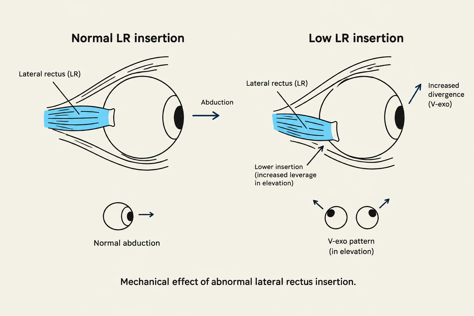
Diagram illustrating the mechanical effect of abnormal lateral rectus insertion on eye movement, comparing normal and low insertion points and their impact on abduction and divergence.
The mechanical advantage of a muscle is heavily dependent on its insertion point.
Horizontal Muscle Insertions:
V-exo: An LR insertion that is too low increases its mechanical advantage during elevation, causing increased divergence.
V-eso: An MR insertion that is too high causes more slack during elevation and increased mechanical advantage during depression, causing increased convergence.
A-eso: An LR insertion that is too high.
A-exo: An MR insertion that is too low.
Oblique and Vertical Rectus Insertions:
V-eso: An SO insertion that is too anterior weakens its abducting force, leading to relatively more convergence in depression.
V-exo: An IO insertion that is too posterior strengthens its abducting force, leading to more divergence in elevation.
Vertical Rectus Shifts: Nasal or temporal shifts in IR or SR insertions alter the adduction/abduction ratios during vertical gaze. For example, a nasal SR insertion can increase adduction during elevation, contributing to an A-eso.
5. Sagittalisation and Muscle Pulleys
Sagittalisation refers to a disruption in the normal parallelism of the oblique muscles. If the angle of origin or insertion is altered, torsion occurs. The eye must then compensate for this cyclo-deviation by altering the contraction of vertical muscles, which induces the A or V pattern.
Muscle Pulleys are connective tissue mechanical origins that guide the extraocular muscles. Mislocation of these pulleys can cause pattern strabismus even without primary oblique dysfunction.
A patterns are associated with incyclorotation of the pulley array.
V patterns are associated with excyclorotation of the pulley array.
Underlying Syndromes and Anomalies
Certain craniofacial conditions are highly associated with pattern strabismus due to underlying orbital anomalies. These include:
Craniosynostosis: Such as Crouzon, Apert, and Pfeiffer syndromes.
Plagiocephaly.
Down Syndrome: Often associated with A-pattern esotropia.
Summary Table of Aetiology
Pattern
Primary Associated Dysfunction
V-eso
SO under-action, IO over-action, MR insertion too high, SO insertion too anterior
V-exo
SR under-action, IO over-action, LR over-action, LR insertion too low, IO insertion too posterior
A-eso
IO under-action, SO over-action, LR under-action, LR insertion too high, IO insertion too anterior
A-exo
IR under-action, SO over-action, MR over-action, MR insertion too low, SO insertion too posterior
Exam Tip: Surgical Principles
When planning surgery for pattern strabismus, the choice of procedure depends on the presence of vertical muscle dysfunction:
If significant vertical muscle over/under-action is present →Operate on vertical muscles.
If vertical muscle action is normal or mild →Operate on horizontal muscles.
Keep reading for free.
Create an account to finish this reading and make it part of your study routine.