


Tuberculosis (TB) remains a significant global public health challenge. In 2025, it was identified as the world's leading cause of death from a single infectious agent. Understanding the distinction between latent infection and active disease is critical for both clinical management and public health control. This article provides a comprehensive overview of the etiology, pathophysiology, diagnostic pathways, and resistance patterns associated with Mycobacterium tuberculosis.
Etiology and Pathophysiology
The disease is caused by Mycobacterium tuberculosis, an aerobic, non-motile, and slow-growing bacillus. Unlike many other bacteria that replicate every 30 minutes, M. tuberculosis replicates approximately once every 24 hours. A defining characteristic of this organism is its complex, waxy cell wall, which is rich in mycolic acids and arabinogalactan. This high lipid content makes the organism impermeable to standard Gram staining; instead, clinicians must use a specialized Ziehl-Neelsen stain. Because these bacteria resist decolorization by acidic alcohol, they are referred to as acid-fast bacilli (AFB).
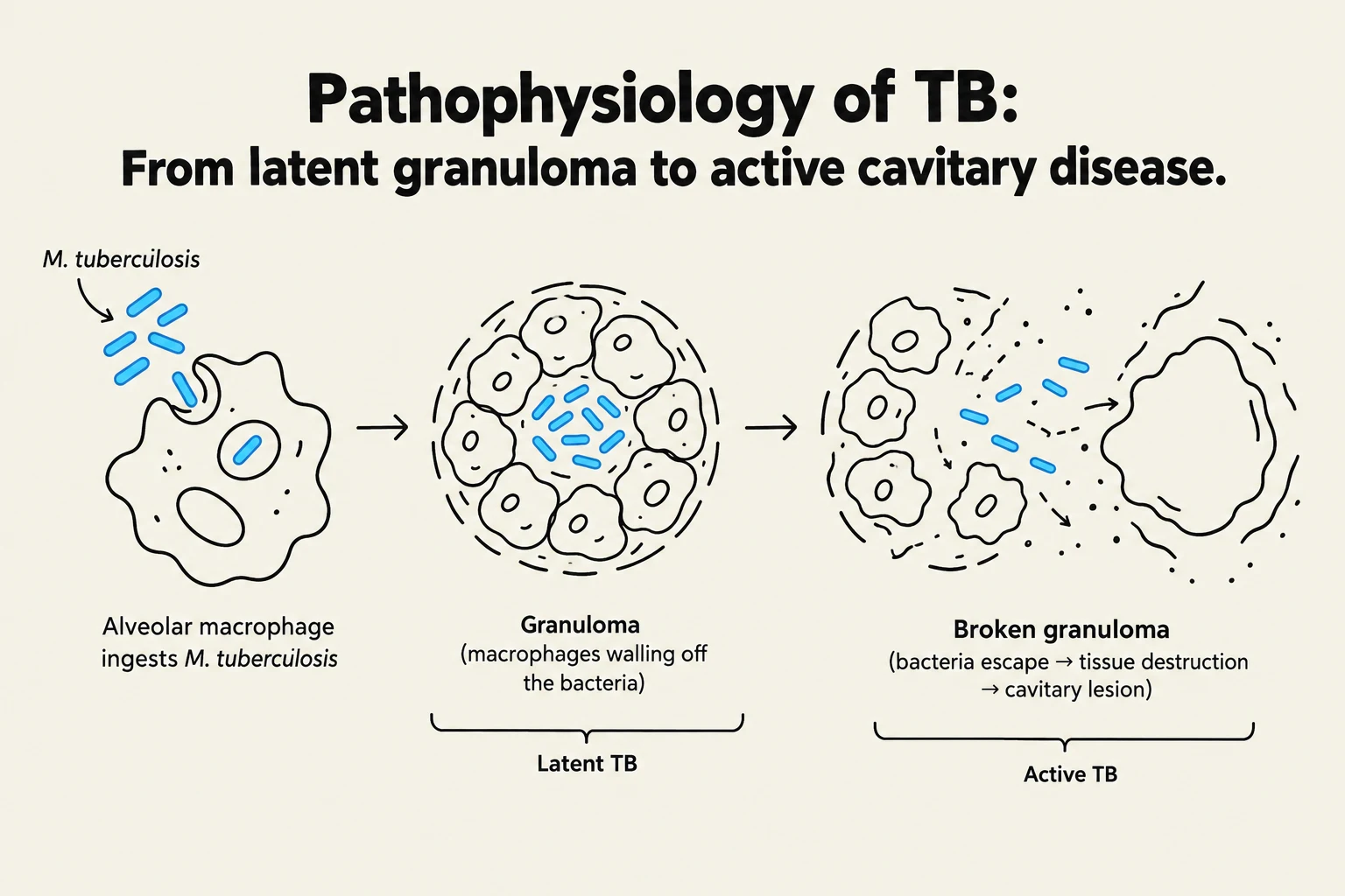
Transmission occurs via respiratory droplets () expelled through coughing, sneezing, or speaking. When these droplets reach the alveoli, they are ingested by alveolar macrophages. While macrophages are the first line of defense, they cannot kill the organism until they are activated by helper T-cells. This activation involves the release of interferon- and tumor necrosis factor (TNF)-.
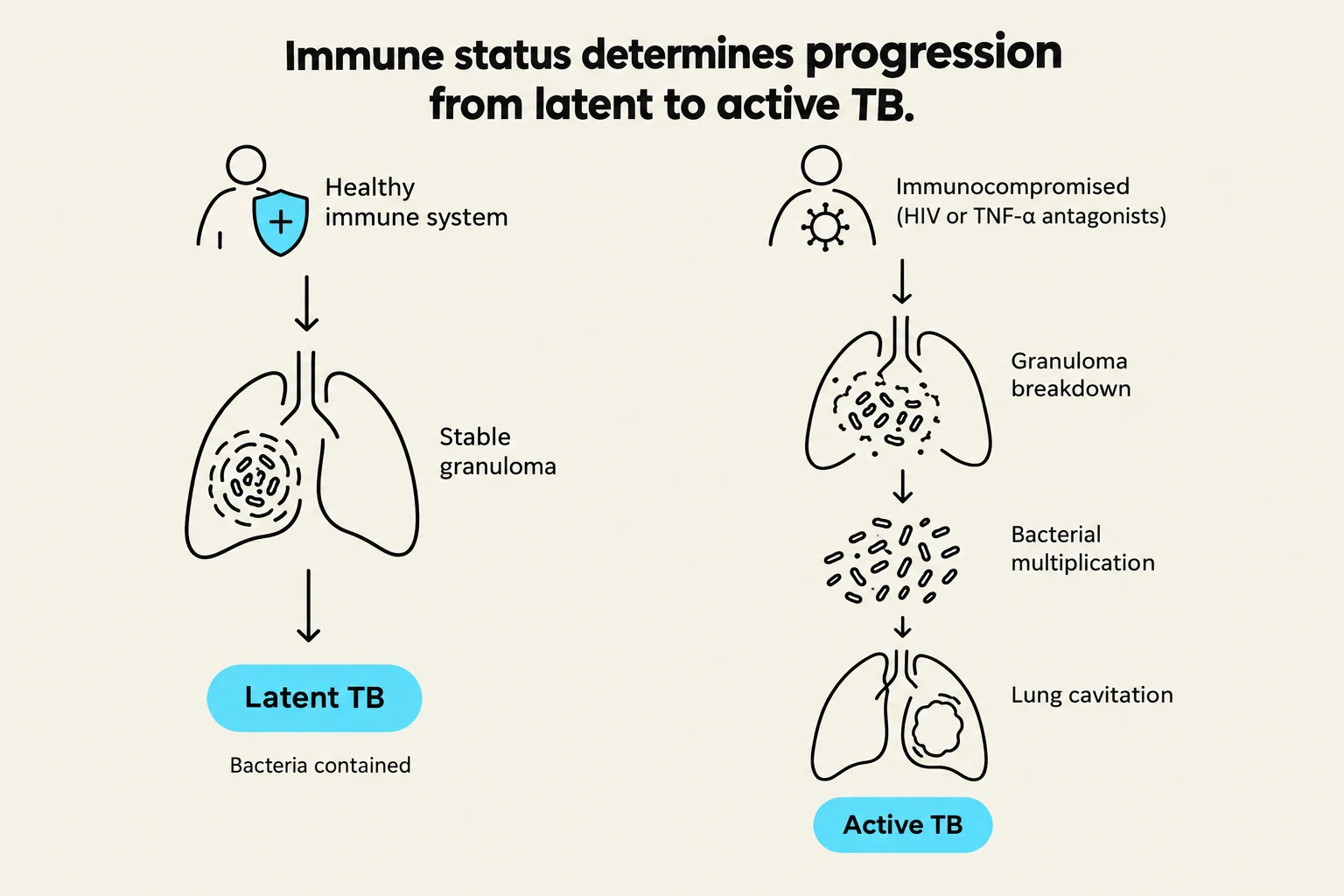
In a healthy immune response, activated macrophages may accumulate around a lesion to form a granuloma, effectively "walling off" the infection. When cell-mediated immunity is intact, the infection is contained, leading to a latent state. However, if the immune system is compromised, the organism can replicate until macrophages burst, potentially creating cavitary lesions in the lungs and allowing the bacteria to spread to the bloodstream and other organs.
Keep reading for free.
Create an account to finish this reading and make it part of your study routine.
Continue reading freeAlready have an account? Log in
Updated