


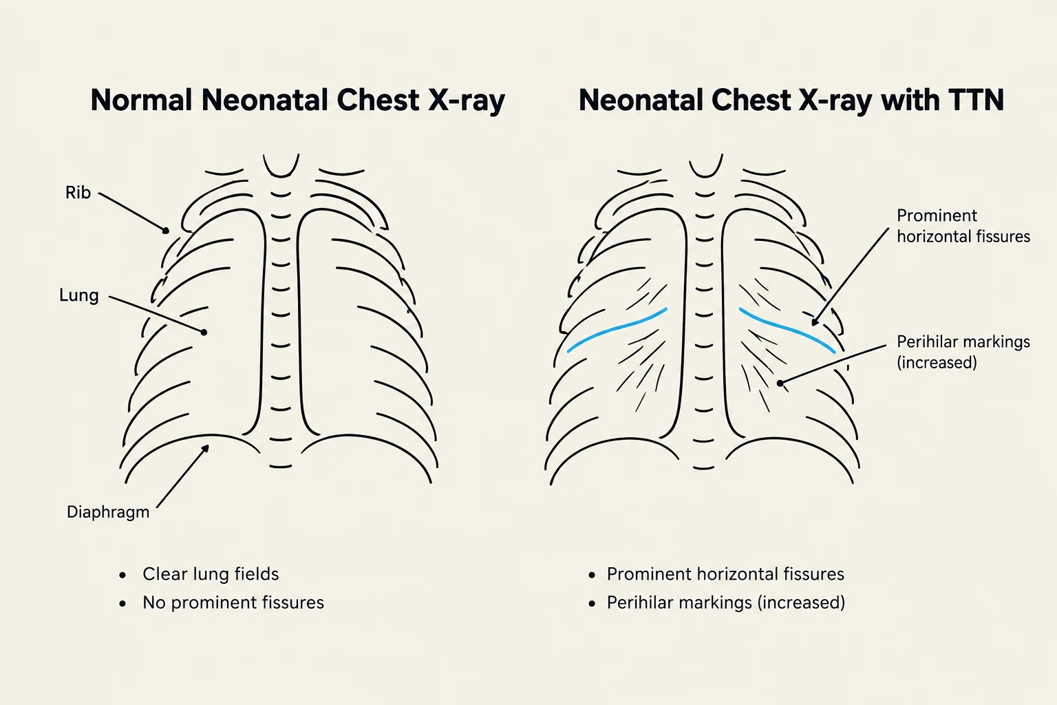
Transient Tachypnoea of the Newborn (TTN) is one of the most common causes of respiratory distress in the neonatal period. While it can be alarming to clinicians and parents due to the rapid breathing observed in the infant, it is fundamentally a self-limiting condition. Understanding the pathophysiology, identifying the specific risk factors, and recognizing the classic radiological patterns are essential skills for any student of neonatology.
Pathophysiology: The Delayed Clearance of Lung Fluid
To understand TTN, one must first understand the transition from intrauterine to extrauterine life. During pregnancy, the fetal lungs are filled with specialized lung fluid that is actively secreted by the pulmonary epithelium. As labor approaches, particularly during a vaginal delivery, several physiological changes occur to prepare the lungs for air breathing.
The most critical change is the switch from fluid secretion to fluid absorption. Hormonal shifts (such as the surge in catecholamines and glucocorticoids) trigger the lung epithelium to stop secreting fluid and start actively absorbing it via sodium channels. In a normal delivery, this process is rapid, allowing the alveoli to expand with air almost immediately.
In TTN, this transition is delayed. The lung fluid remains within the interstitial and alveolar spaces, interfering with efficient gas exchange and causing the infant to breathe rapidly to compensate for the decreased lung compliance.
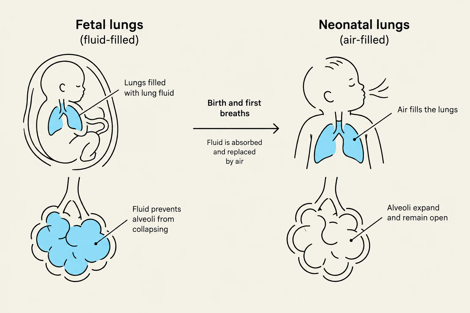
Exam Tip: When asked about the mechanism of TTN, look for keywords like "delayed clearance of fetal lung fluid" or "impaired alveolar fluid absorption." Avoid confusing this with Respiratory Distress Syndrome (RDS), which is caused by surfactant deficiency rather than fluid retention.

Keep reading for free.
Create an account to finish this reading and make it part of your study routine.
Continue reading freeAlready have an account? Log in
Updated