Tracheostomy Care and Respiratory Complications Management
A comprehensive guide to tracheostomy procedures, including assessment, maintenance, safety protocols, and suctioning techniques. It also covers the management of aspiration, pneumonia, pleural disorders, chest tubes, atelectasis, and mechanical ventilation.
Effective respiratory management is a cornerstone of critical care, particularly for patients with compromised airways or impaired gas exchange. This article explores the complexities of tracheostomy care, the management of pleural disorders, and the prevention and treatment of atelectasis and other significant respiratory complications.
Tracheostomy: Procedure and Safety Protocols
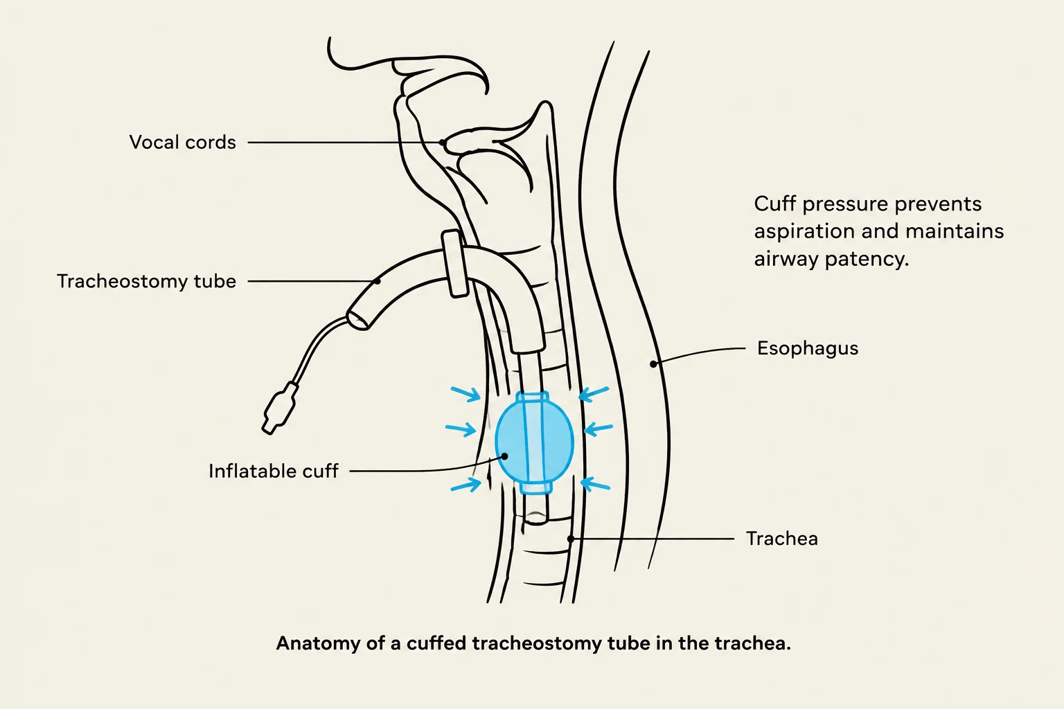
Anatomy of a cuffed tracheostomy tube in the trachea.
A tracheostomy is a surgical procedure that creates an opening into the trachea to insert an indwelling tracheostomy tube. This intervention is indicated for patients who require prolonged mechanical ventilation, need to bypass an upper airway obstruction, require assistance with secretion removal, or have suffered complications such as paralyzed vocal cords.
Assessment and Maintenance
When assessing a patient with a tracheostomy, clinicians must identify the tube size and determine if the tube is cuffed or uncuffed. If a cuff is present, maintaining appropriate pressure is vital to prevent both airway trauma and aspiration. Cuff pressure should be maintained between 20 to 25 cm H2O and must be checked every eight hours. To prevent the airway from drying out, warm humidification should be utilized.
Bedside Safety Essentials
Because a tracheostomy represents a high-risk airway, specific emergency equipment must be kept at the patient's bedside at all times:
An extra tracheostomy tube of the same size as the current tube.
An extra tracheostomy tube one size smaller.
An obturator.
Suction equipment and dressing kits.
Suctioning Techniques
Suctioning is performed as needed to maintain airway patency. To ensure patient safety during this procedure, clinicians must follow strict protocols:
Hyperoxygenate: Provide the patient with 100%FiO2 for one minute prior to suctioning.
Insertion: Advance the catheter until resistance is met or the patient coughs.
Withdrawal: Withdraw the catheter with intermittent suctioning and a rotating motion.
Duration and Frequency: Each suction pass must not exceed 15 seconds, and there should be a maximum of three attempts per session.
Warning: Complications
Improper tracheostomy management can lead to severe complications, including air embolism, aspiration, subcutaneous emphysema (air leaking into subcutaneous tissue), laryngeal damage, tube dislodgement, and accidental decannulation.
Aspiration and Pneumonia
Aspiration occurs when foreign material is inhaled into the lungs. This is a significant risk factor for developing aspiration pneumonia, a condition where the inhaled material acts as a reservoir for bacteria.
Risk Factors and Prevention
Patients at risk for aspiration often exhibit swallowing difficulties, such as coughing during intake or "pocketing" food in the cheeks, which may indicate weakened muscles. To prevent aspiration, nursing interventions include:
Elevating the head of the bed to >30∘.
Verifying the placement of G-tubes before feedings.
Transitioning patients to thickened liquids if they struggle with thin liquids.
Pneumonia is an inflammation of the lung parenchyma caused by bacteria, viruses, or fungi. Symptoms often include a sudden onset of chills, fever, pleuritic chest pain, and the production of purulent sputum. Influenza is a common viral precursor; antivirals like oseltamivir are most effective when initiated within 24–48 hours of symptom onset.
Pleural Disorders and Chest Tubes
Pleural effusion is the accumulation of fluid in the pleural space, often secondary to heart failure, malignancy, or infection. Diagnosis is typically achieved through chest X-ray, CT scans, or thoracentesis.
Pneumothorax
A major complication of thoracentesis is pneumothorax, which is the presence of air in the pleural space. This can be spontaneous or traumatic (e.g., from an accident). Clinical manifestations include dyspnea, tachycardia, asymmetrical chest movement, and potentially tracheal deviation in severe cases.
Chest Tube Management
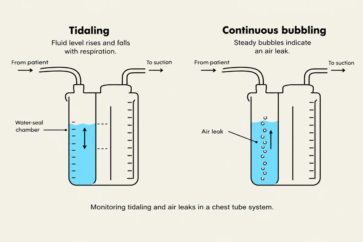
Diagrams illustrating tidaling and continuous bubbling in a chest tube system, indicating normal respiration and an air leak, respectively.
Chest tubes are inserted into the pleural space to drain air, blood, or fluid to facilitate lung expansion. Nurses must monitor the following:
Tidaling: The expected fluctuation of fluid in the water-seal chamber with respiration. The cessation of tidaling may indicate the lung has re-expanded or there is an obstruction (like a kink).
Continuous Bubbling: In the water-seal chamber, continuous bubbling indicates an air leak in the system. Note that intermittent bubbling during coughing or sneezing is expected when draining air.
Emergency Protocol: Accidental Removal
If a chest tube is accidentally removed from the chest, cover the site immediately with a sterile gauze dressing and tape it on three sides. This creates a one-way valve that allows air to escape during exhalation but prevents it from entering during inhalation.
Atelectasis and Respiratory Support
Comparison of collapsed alveoli versus expanded alveoli.Atelectasis is the collapse or closure of the alveoli. It is common in postoperative settings and can be acute or chronic. Symptoms include increased respiratory effort, hypoxemia (low blood oxygen), dyspnea (shortness of breath), and decreased breath sounds or crackles upon auscultation.
Management Strategies
To prevent and manage atelectasis, clinicians utilize:
Incentive Spirometry: Encouraging deep breathing to expand the lungs.
Frequent Turning and Early Mobilization: To prevent stasis and promote lung expansion.
Positive End-Expiratory Pressure (PEEP): Providing pressure during expiration to keep alveoli open and treat hypoxia.
Mechanical Ventilation and Respiratory Failure
Mechanical ventilation is used to support patients experiencing respiratory failure, which is the inability to adequately ventilate or oxygenate.
Ventilator Modes
PEEP: Delivers preset pressure during expiration to treat persistent hypoxia.
Pressure Support: Helps keep alveoli from collapsing and decreases the work of breathing.
Critical care for ventilated patients includes performing oral care with chlorhexidine every 2 hours to prevent Ventilator-Associated Pneumonia (VAP). Medications such as sedatives (e.g., Versed) and analgesics (e.g., Fentanyl) are used to prevent the patient from "fighting" the ventilator.
Note on Paralytics: Neuromuscular blocking agents (paralytics) only paralyze muscles; they provide no sedation or pain relief and must always be used in conjunction with sedation and analgesia.
Weaning and Complications
When weaning a patient from a ventilator, clinicians must monitor for distress. Signs that a patient is not tolerating weaning include:
Respiratory rate >30 breaths/min.
SpO2<90%.
Heart rate or blood pressure changes >20%.
Increased restlessness, anxiety, or decreased level of consciousness.
Complications of mechanical ventilation include barotrauma (pressure-related injury), volutrauma (volume-related injury), oxygen toxicity, and hemodynamic compromise due to increased thoracic pressure decreasing venous return.
Keep reading for free.
Create an account to finish this reading and make it part of your study routine.