An overview of the thyroid gland's role in the endocrine system, covering its anatomy, hormone biosynthesis, the hypothalamic-pituitary-thyroid axis, and the physiological roles of T4 and T3. It also discusses clinical correlations like hyperthyroidism and hypothyroidism.
The thyroid gland is a vital component of the endocrine system, acting as a central regulator of the body's metabolic rate. Located in the lower anterior neck, this butterfly-shaped gland consists of two lobes connected by a central band known as the isthmus. While it is most famous for producing hormones that govern energy expenditure, the thyroid and its associated structures play diverse roles in development, thermogenesis, and even calcium regulation.
Anatomy and Cellular Structure
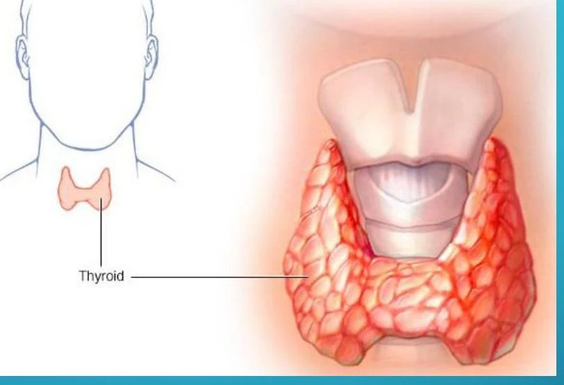
The thyroid gland is positioned in front of the trachea.
An illustration of the human thyroid gland, located in the neck.
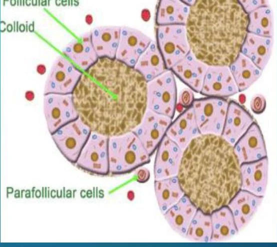
To understand how the gland functions, we must look at its microscopic organization. The thyroid is composed of functional units called follicles. These follicles are lined by thyroid follicular cells and filled with a protein-rich fluid called colloid.
Beyond the follicular cells, there are specialized cells known as parafollicular C cells. These cells are responsible for secreting calcitonin, a hormone involved in calcium homeostasis. While the parathyroid glands (four small "dots" located on the posterior surface of the thyroid) are the primary regulators of serum calcium, calcitonin provides an additional layer of control.
Diagram of thyroid follicles showing follicular cells, colloid, and parafollicular cells.
Thyroid Hormone Biosynthesis
The production of thyroid hormones is a complex biochemical process that occurs within the follicular cells and requires a steady supply of iodine. Iodine is an essential micronutrient for this process; an intake of less than 50 mcg daily can lead to hypothyroidism.
The biosynthesis involves several steps, including the trapping of iodine, the synthesis of a protein called thyroglobulin, and the subsequent iodination of tyrosine residues on that protein.
The Hypothalamic-Pituitary-Thyroid (HPT) Axis
Thyroid hormone production is not autonomous; it is tightly regulated by a multi-tiered feedback system known as the Hypothalamic-Pituitary-Thyroid (HPT) axis.
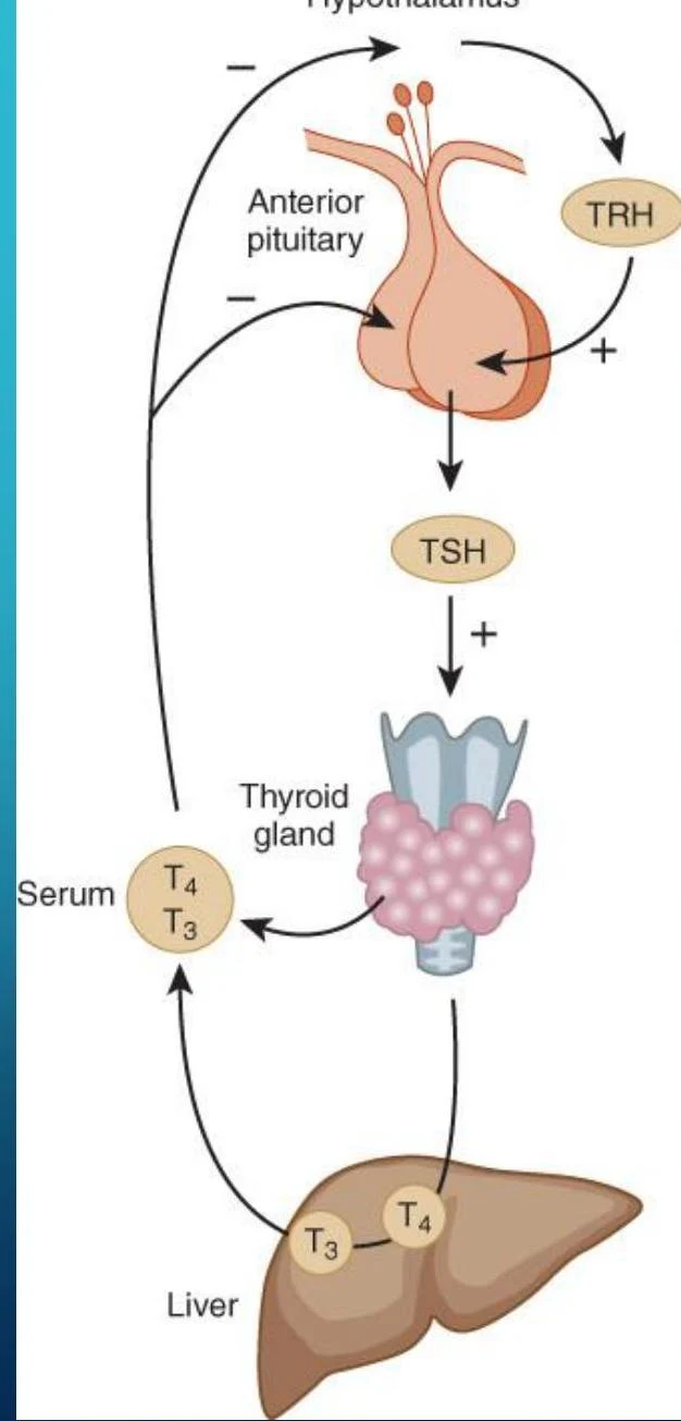
The Hypothalamus: When thyroid hormone levels in the blood drop, the hypothalamus releases Thyrotropin-Releasing Hormone (TRH).
The Anterior Pituitary: TRH travels to the anterior pituitary, stimulating it to manufacture and release Thyroid Stimulating Hormone (TSH).
The Thyroid Gland: TSH enters the circulation and acts on the thyroid follicular cells, prompting them to produce and release thyroid hormones (T3 and T4).
Negative Feedback Loops
The system is self-correcting through negative feedback. Once the levels of T3 and T4 in the bloodstream reach an adequate threshold, these hormones signal back to the hypothalamus and the anterior pituitary to inhibit the release of TRH and TSH, respectively. This prevents the overproduction of hormones and maintains metabolic stability.
This diagram illustrates the hormonal regulation of the thyroid gland. The hypothalamus releases TRH, which stimulates the anterior pituitary to release TSH. TSH then acts on the thyroid gland to produce T4 and T3. These hormones are then released into the serum and affect the liver, which also plays a role in hormone metabolism.
T4 and T3: Roles and Transport
The two primary thyroid hormones are Thyroxine (T4) and Tri-iodothyronine (T3). The fundamental difference between them is the number of iodine molecules attached to the thyroglobulin base.
T4 (Thyroxine): Often considered the "pro-hormone," it is produced in larger quantities.
T3 (Tri-iodothyronine): The more biologically active form. Much of the T3 used by the body is created by the de-iodination of T4 in peripheral tissues.
Physiological Roles
T3 is the primary driver of metabolism and development. Its functions include:
Influencing tissue growth and brain maturation.
Increasing body heat production (thermogenesis) and oxygen consumption.
Increasing the number of beta-adrenergic receptors, which affects how the body responds to adrenaline.
Protein Binding and Transport
In the bloodstream, thyroid hormones exist in two states: unbound (free) and bound.
Unbound: Only a tiny fraction (0.04% of T4 and 0.4% of T3) is "free" and available for immediate cellular activity.
Bound: The vast majority (>99.9%) is bound to carrier proteins, which act as a reservoir. The primary carriers are Thyroxine-binding globulin (TBG), Thyroxine-binding prealbumin (TBPA), and albumin.
Clinical Note: Because most hormone is protein-bound, any alteration in binding proteins (like TBG) can cause abnormal total T4 or T3 levels in lab tests, even if the "free" active hormone levels remain normal.
Clinical Correlations: Hyperthyroidism and Hypothyroidism
When the HPT axis or the thyroid gland itself malfunctions, it results in clinical disorders.
Hypothyroidism (Low Metabolic Activity)
Hypothyroidism occurs when there is insufficient thyroid hormone. It is often categorized by the location of the defect:
Primary Hypothyroidism: The defect is in the thyroid gland itself (e.g., Hashimoto's thyroiditis, where auto-antibodies destroy the gland).
Secondary Hypothyroidism: The defect is in the anterior pituitary (e.g., hypopituitarism).
Tertiary Hypothyroidism: The defect is in the hypothalamus.
Common Signs: Bradycardia (slow heart rate), cold intolerance, weight gain, constipation, and delayed reflex relaxation.
Hyperthyroidism occurs when there is an excess of thyroid hormone. The most common cause is Graves' Disease, an autoimmune disorder where antibodies stimulate the TSH receptors (TRAb), causing the gland to overproduce hormone.
Common Signs: Tachycardia (fast heart rate), heat intolerance, weight loss, tremors, and in Graves' disease, specific symptoms like ophthalmopathy (bulging eyes).
Condition
Pathogenic Mechanism
TSH Level
RAIU (Uptake)
Graves' Disease
Anti-TSHR antibodies
↓
↑
Toxic Adenoma
Benign nodule
↓
↑
Thyroiditis
Hormone leakage
↓
↓
Exogenous Hormone
Excess medication
↓
↓
Table 14.4: Disorders Associated with Thyrotoxicosis
Exam Tip: When interpreting thyroid labs, always look at TSH first. TSH is the most sensitive indicator of thyroid function. In primary hypothyroidism, TSH will be high (trying to stimulate the failing gland), whereas in primary hyperthyroidism, TSH will be low (suppressed by the excess hormone).
Keep reading for free.
Create an account to finish this reading and make it part of your study routine.