An overview of the thyroid gland's anatomy, hormone synthesis, the hypothalamic-pituitary-thyroid axis, and diagnostic methods for thyroid disorders like hypothyroidism and hyperthyroidism.
The thyroid gland is a butterfly-shaped endocrine organ located in the lower anterior neck, bridging the two lobes across the trachea. It plays a critical role in regulating the body's metabolic rate, influencing everything from heat production and oxygen consumption to brain maturation and tissue growth. Because thyroid hormones affect nearly every organ system, clinical assessment of thyroid function is a cornerstone of metabolic medicine.
Anatomy and Hormone Synthesis
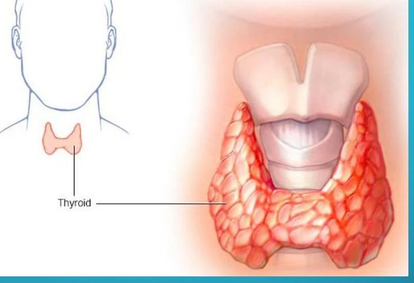
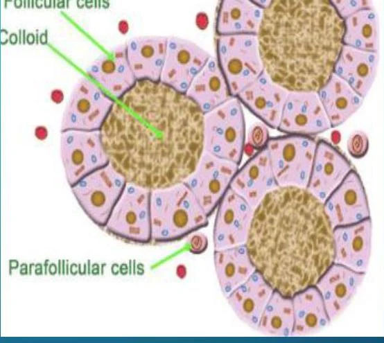
The thyroid gland consists of two lobes and an isthmus. Within the gland, thyroid follicular cells are responsible for manufacturing thyroid hormones.
An illustration of the human thyroid gland, located in the neck.
Diagram of thyroid follicles showing follicular cells, colloid, and parafollicular cells.
The synthesis of these hormones requires iodine (I2); an intake of less than 50 mcg daily can lead to hypothyroidism. While the follicular cells produce the primary thyroid hormones, the parafollicular C cells secrete calcitonin, which is involved in calcium homeostasis.
The Hypothalamic-Pituitary-Thyroid Axis
Thyroid hormone production is governed by a negative feedback loop known as the hypothalamic-pituitary-thyroid axis.
Hypothalamus: When thyroid hormone levels are low, the hypothalamus releases Thyrotropin-Releasing Hormone (TRH).
Anterior Pituitary: TRH stimulates the anterior pituitary to manufacture and release Thyroid Stimulating Hormone (TSH).
Thyroid Gland: TSH acts on the thyroid to stimulate the production and release of Thyroxine (T4) and Triiodothyronine (T3).
Once T3 and T4 levels reach adequate concentrations in the blood, they signal the hypothalamus to stop releasing TRH, completing the feedback loop.
Thyroid Hormones: T3 and T4
There are two primary medically important thyroid hormones:
Thyroxine (T4): Often considered the prohormone, it is the more abundant form in circulation.
Triiodothyronine (T3): The more active form. T4 is de-iodinated in peripheral tissues to become T3.
In the bloodstream, these hormones exist in two states: Bound and Unbound (Free). Over 99.9% of total T4 and 99.6% of T3 are bound to protein carriers such as Thyroxine-binding globulin (TBG), thyroxine-binding prealbumin (TBPA), and albumin. Only the "free" portion is biologically available for cellular activity.
Diagnostic Evaluation of Thyroid Function
TSH and Free Hormone Testing
TSH is the most useful single test for assessing thyroid function. Because of the feedback loop, TSH levels often change in the opposite direction of thyroid hormones.
Result Pattern
Interpretation
High TSH + Low Free T4
Primary Hypothyroidism
Low TSH + High Free T4
Primary Hyperthyroidism (Thyrotoxicosis)
Low TSH + Low Free T4
Secondary Hypothyroidism or Nonthyroidal Illness
Exam Tip: Always consider "Nonthyroidal Illness" (Euthyroid Sick Syndrome) when a critically ill patient presents with low TSH, T3, and T4. This is a physiological adaptation to severe systemic illness rather than a primary thyroid disorder.
Antibody Testing
Autoimmune processes are common causes of thyroid dysfunction. Specific antibodies can help differentiate between them:
Thyroperoxidase (TPO) Antibody: Plays a role in the pathogenesis of hypothyroidism and is often positive in Hashimoto's.
Thyroglobulin Antibody (anti-Tg): Associated with autoimmune destruction or inflammation; it may leak into the blood during thyroiditis.
Thyrotropin Receptor Antibody (TRAb): These stimulating antibodies are the hallmark of Graves' disease, as they mimic TSH and cause the gland to overproduce hormone.
Nuclear Medicine and Imaging
Radioactive Iodine Uptake (RAIU): This test assesses the metabolic activity of the thyroid. High uptake indicates an overactive gland (e.g., Graves' disease), while low uptake suggests inflammation or exogenous hormone ingestion.
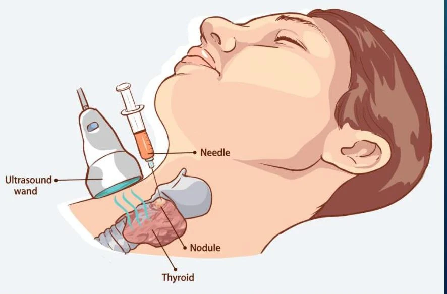
Thyroid Ultrasound: Highly effective at detecting small nodules (less than 1 cm).
Fine-Needle Aspiration (FNA): The most accurate tool for evaluating thyroid nodules. It uses a thin needle to obtain cells for cytologic exam, helping to avoid unnecessary surgery in benign cases.
Illustration of an ultrasound-guided needle biopsy of a thyroid nodule.
Clinical Disorders
Hypothyroidism
Hypothyroidism is defined by low free T4 levels.
Primary Hypothyroidism: The defect is in the thyroid gland itself (e.g., Hashimoto's Thyroiditis, where auto-antibodies destroy the gland).
Secondary/Tertiary: The defect lies in the pituitary or hypothalamus.
Common Symptoms: Cold intolerance, weight gain, bradycardia (slow heart rate), constipation, and depression.
Thyrotoxicosis (Hyperthyroidism)
Thyrotoxicosis refers to the clinical state of excess thyroid hormone in peripheral tissues.
Graves' Disease: The most common cause of hyperthyroidism. It is an autoimmune disorder where TRAbs stimulate the TSH receptor. It is characterized by thyrotoxicosis, goiter, and unique signs like ophthalmopathy (bulging eyes) and dermopathy.
Subacute Thyroiditis: Characterized by transient changes in hormone levels due to inflammation. As the inflammation subsides, stored hormone leaks into the blood, often causing temporary thyrotoxicosis followed by hypothyroidism.
Clinical Note: When evaluating a patient with a thyroid nodule, remember that while nodules are common, only about 5-9% of nonpalpable nodules are actually cancerous.
Keep reading for free.
Create an account to finish this reading and make it part of your study routine.