A comprehensive guide to assessing the thorax and lungs, covering anatomy, subjective data collection, and objective examination techniques including inspection, palpation, percussion, and auscultation.
The respiratory system is vital for life, facilitating the exchange of oxygen and carbon dioxide between the atmosphere and the blood. A thorough assessment of the thorax and lungs requires a blend of anatomical knowledge, precise history-taking, and a systematic physical examination. By understanding the relationship between the structural integrity of the thoracic cage and the physiological function of the lungs, clinicians can effectively identify deviations from health, such as airway obstruction, infection, or impaired gas exchange.
Anatomy of the Thoracic Cage and Lungs
The thoracic cage serves as a protective shield for the heart and lungs while providing the structural framework necessary for breathing. It is composed of the sternum—which includes the manubrium, the body, and the xiphoid process—along with 12 pairs of ribs, 12 thoracic vertebrae, costal cartilages, and the intercostal muscles.
When performing an assessment, clinicians rely on specific landmarks to orient themselves. These include the suprasternal notch, the sternal angle (Angle of Louis) located at the second rib, and the costal angle.
The lungs themselves are asymmetrical to accommodate the body's internal organs. The right lung is larger, consisting of three lobes (upper, middle, and lower) divided by horizontal and oblique fissures. In contrast, the left lung has only two lobes (upper and lower) to make room for the heart. The lung apices extend above the clavicles, while the bases rest upon the diaphragm.
Diagram illustrating the human rib cage and lungs, highlighting the lobes of the right and left lungs and noting the asymmetry due to the heart's position.
The process of breathing is regulated by the respiratory center in the brainstem. This center is highly sensitive to chemical changes in the blood, primarily responding to fluctuations in CO2 levels and pH. Inspiration occurs when the diaphragm contracts and descends, increasing thoracic volume and lowering pressure to draw air in; expiration at rest is a passive process driven by the elastic recoil of the lungs.
Subjective Data Collection: The Patient History
Before touching the patient, the clinician must gather subjective data to understand the context of the patient's respiratory health. This involves asking about cough (onset, duration, sputum color, or presence of blood), dyspnea (shortness of breath), and chest pain. It is also critical to investigate lifestyle and environmental factors, such as smoking or vaping habits, secondhand smoke exposure, and occupational risks like dust, chemicals, or mold.
To ensure a comprehensive characterization of symptoms, clinicians use the PQRST(A)U mnemonic:
P (Provocation/Palliation): What triggers the symptom (e.g., exercise, lying flat) or makes it better?
Q (Quality): What is the character? (e.g., Is the cough dry or productive? Is the pain sharp or burning?)
R (Region/Radiation): Where is the discomfort located, and does it spread?
S (Severity): On a scale of 0–10, how intense is the pain? How much does it limit activity?
T (Timing): When did it start? Is it constant, or does it occur only at night?
A (Associated symptoms): Are there other signs like fever, wheeze, cyanosis, or dizziness?
U (Understanding): What does the patient believe is causing the symptom?
Exam Tip: When assessing dyspnea, always ask about orthopnea (difficulty breathing while lying flat) and paroxysmal nocturnal dyspnea (sudden attacks at night), as these can be significant indicators of cardiac involvement.
Objective Assessment: The IPPA Sequence
The physical examination follows the systematic IPPA sequence: Inspection, Palpation, Percussion, and Auscultation.
1. Inspection
The goal of inspection is to observe the "work of breathing." The clinician should assess the respiratory rate, rhythm, and depth. Key observations include:
Symmetry: Does the chest expand equally on both sides?
Chest Shape: The normal Anteroposterior (AP) to transverse diameter should be approximately 1:2.
Effort: Is the patient using accessory muscles to breathe?
Skin Color: Look for cyanosis (bluish discoloration) or clubbing of the fingernails, which may indicate chronic hypoxemia.
2. Palpation
Palpation is used to confirm findings from inspection and to detect abnormalities beneath the skin.
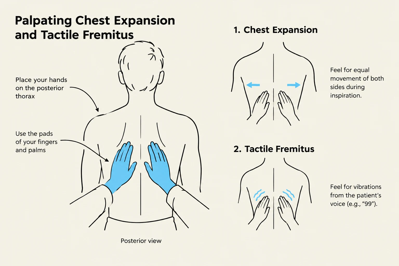
Chest Expansion: Place your hands on the posterior lower chest (at the level of T9–T10). As the patient inhales deeply, your thumbs should move apart symmetrically.
Tactile Fremitus: Use the ulnar edge of your hand to feel for vibrations while the patient repeats the phrase "ninety-nine." Compare both sides for symmetry.
Other findings: Check for tenderness, masses, or crepitus (a crunchy sensation caused by air under the skin).
This illustration demonstrates the medical techniques for palpating chest expansion and tactile fremitus on the posterior thorax.
3. Percussion
Percussion involves tapping on the intercostal spaces to assess the density of the underlying lung tissue. The clinician should move side-to-side from the apices to the bases, avoiding the scapulae.
Resonance: The normal sound heard over air-filled lung tissue.
Dullness: Indicates fluid or consolidation (e.g., pneumonia or pleural effusion).
Hyperresonance: Indicates excess air, often seen in conditions like emphysema.
4. Auscultation
The final step is auscultation using the diaphragm of the stethoscope on bare skin. The patient should breathe deeply through their mouth. Clinicians must compare corresponding points from the apices to the bases to identify normal or adventitious sounds.
Sound Type
Description
Common Association
Vesicular
Soft, low-pitched; inspiration > expiration
Normal peripheral lung fields
Bronchovesicular
Medium pitch; inspiration ≈ expiration
Major bronchi
Crackles (Rales)
Discontinuous popping sounds
Fluid, pneumonia, or collapsed airways
Wheezes
Continuous musical, high-pitched sounds
Narrowed airways (Asthma/COPD)
Stridor
Harsh, high-pitched inspiratory sound
Emergency: Upper airway obstruction
Clinical Alert: If you hear stridor, this is a medical emergency suggesting a blocked upper airway. If you hear diminished or silent breath sounds in a patient having an asthma attack, they may be entering respiratory failure.
Keep reading for free.
Create an account to finish this reading and make it part of your study routine.