Explore the anatomy and physiology of the urinary system, including the ureters, bladder, and urethra. Understand the transformation of plasma to urine through filtration, reabsorption, and secretion, and learn about the effects of aging on renal function and urinalysis.
The urinary system is a sophisticated biological filtration and waste management network. Its primary mission is to maintain homeostasis by regulating fluid volume, electrolyte balance, and blood pressure, while simultaneously eliminating metabolic wastes, excess salts, and toxins. To understand how the body transitions from circulating blood to excreting urine, we must examine the specialized organs of storage, the complex chemical evolution of urinary fluids, and the physiological changes that occur as we age.
Organs of Storage and Transport
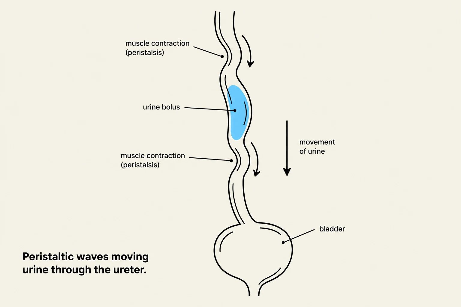
Peristaltic waves moving urine through the ureter.
Once the kidneys have processed the blood, the resulting fluid must be transported, stored, and eventually expelled from the body through a series of specialized structures.
The Ureters
The journey from the kidneys to the bladder is facilitated by the ureters. These are tubes lined with a mucous membrane and composed of smooth muscle. Rather than relying solely on gravity, the ureters utilize peristaltic waves—rhythmic muscular contractions—to actively move urine toward the bladder.
The Urinary Bladder
The urinary bladder acts as a sterile, hollow, muscular reservoir located behind the symphysis pubis. It is designed for flexibility; the bladder walls contain rugae (muscular folds) that allow the organ to distend as it fills, typically holding between 300–500 ml of urine. The floor of the bladder is a specialized triangular region known as the trigone.
Micturition and Control
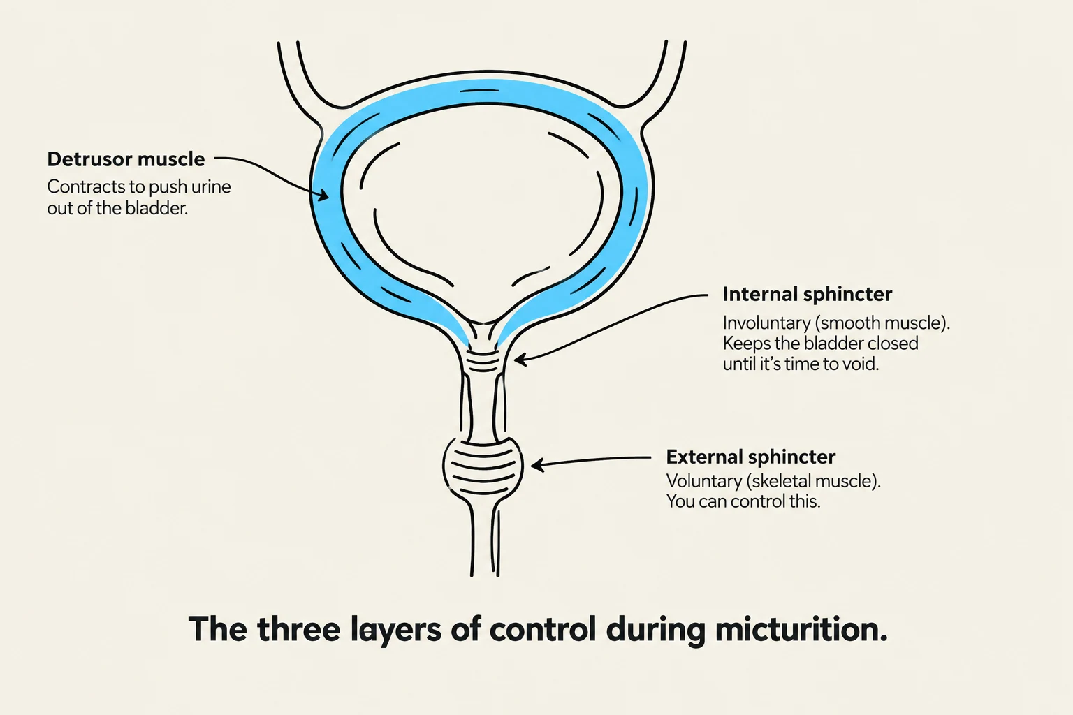
Diagram illustrating the detrusor muscle, internal sphincter, and external sphincter, which are the three layers of control during micturition.
The process of emptying the bladder, known as micturition (or voiding), is a coordinated muscular event. It involves:
The Detrusor Muscle: The muscular layer of the bladder wall that contracts to expel urine.
Internal Sphincter: An involuntary muscle that relaxes automatically during the micturition process.
External Sphincter: A voluntary muscle that must relax to allow urine to pass.
When these processes are not properly coordinated, a person may experience incontinence, the loss of bladder control.
The Urethra
The final pathway is the urethra, the tube that carries urine to the outside of the body via the urinary meatus (or urinary canal). There are significant anatomical differences between the sexes:
In men: The urethra is approximately 8 in long and passes through both the prostate gland and the penis.
In women: The urethra is much shorter, approximately 1.5 in long.
Clinical Note: The shorter length of the female urethra is a significant anatomical factor in the increased risk of urinary tract infections (UTIs) compared to males.
The Chemical Evolution: Plasma to Urine
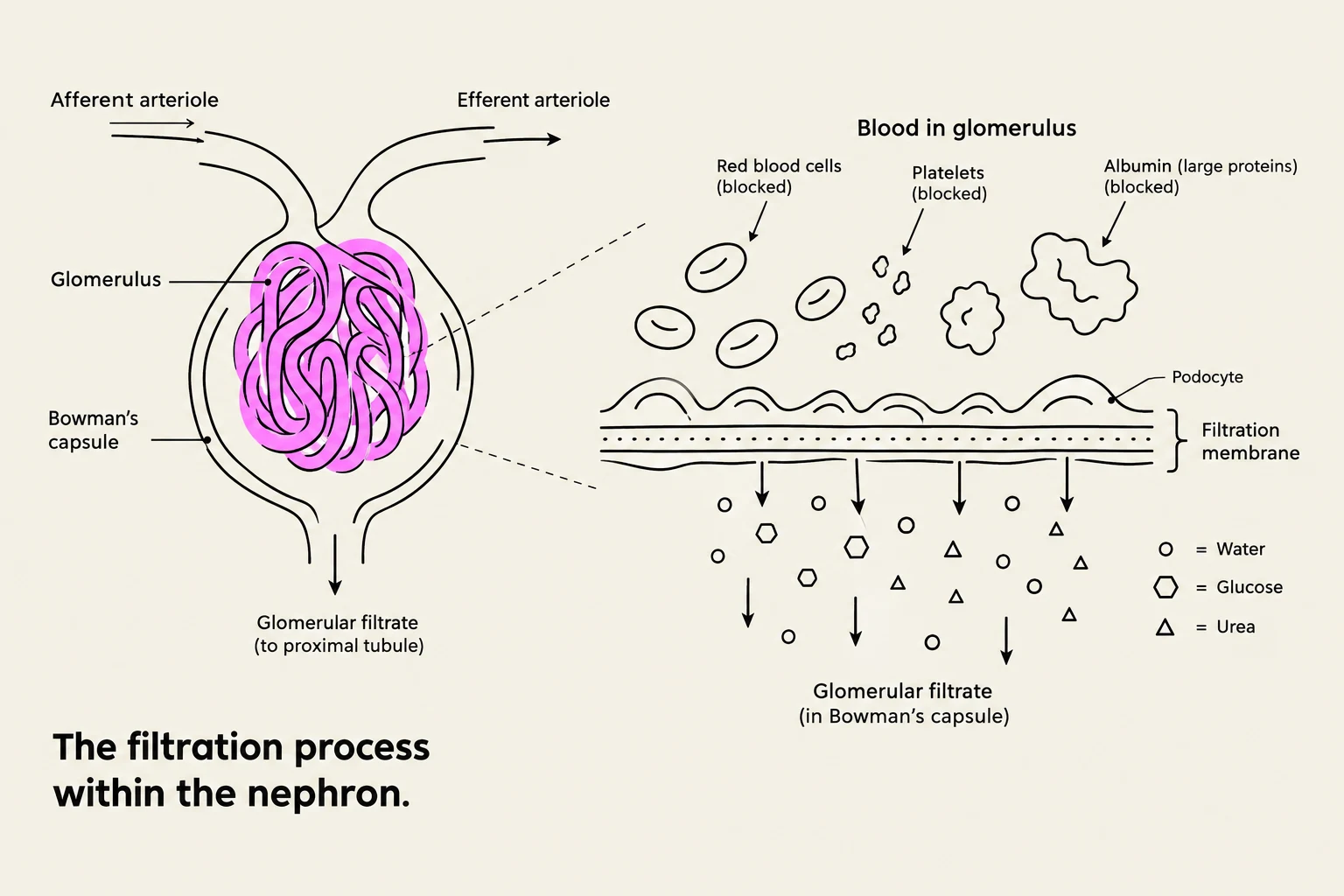
Diagram illustrating the filtration process within the nephron, showing blood flow through the glomerulus and Bowman's capsule, and the filtration of water, glucose, and urea.
A common misconception is that urine is simply "filtered blood." In reality, the fluid undergoes a profound transformation as it moves through the nephron, the functional unit of the kidney.
1. Plasma
Plasma is the liquid portion of the blood. It is a complex mixture containing water, ions, amino acids, vitamins, wastes, and large proteins.
2. Glomerular Filtrate
As blood passes through the glomerulus (a knotted cluster of capillaries), it is filtered into Bowman's capsule. This resulting fluid is the glomerular filtrate. While the filtrate is similar to plasma in that it contains water, salts, electrolytes, glucose, urea, and creatinine, it is distinct because it excludes large substances. Specifically, blood cells, platelets, antibodies, and albumin (a large protein) cannot pass through the filtration membrane.
The body produces a massive amount of filtrate—approximately 200 L per day.
3. Urine
The final product is urine. As the filtrate travels through the proximal convoluted tubule, the Loop of Henle, and the distal tubule, the kidneys perform tubular reabsorption—taking back necessary substances like glucose and water—and tubular secretion—adding extra wastes into the tubule.
The end result is a concentrated fluid containing only the wastes intended for elimination. While 200 L of filtrate is produced daily, normal excretion is typically less than 2 L per day.
Fluid Type
Composition Highlights
Key Exclusions
Plasma
Water, ions, amino acids, vitamins, proteins
N/A
Glomerular Filtrate
Water, glucose, salts, urea, creatinine
RBCs, WBCs, Platelets, Albumin
Urine
Concentrated wastes, excess salts, toxins
Essential nutrients (reabsorbed)
Clinical Assessment: Urinalysis
To evaluate renal health, clinicians perform a urinalysis. This diagnostic tool examines several characteristics:
Physical Properties: Color, appearance, and odor.
Chemical Properties: pH, specific gravity, glucose, ketones, and protein.
Microscopic Examination: The presence of Red Blood Cells (RBCs), White Blood Cells (WBCs), or other cell types.
Specialized Screening: Testing for toxins, drugs, or the hCG hormone (which indicates pregnancy).
Effects of Aging on the Urinary System
As the body ages, the urinary system undergoes physiological declines that can impact quality of life and health stability.
Reduced Nephron Count: There is a natural reduction in the total number of functional nephrons.
Loss of Muscle Tone: The bladder walls lose muscle tone, which can lead to increased urinary frequency.
Inefficient Emptying: If the bladder does not empty completely, residual urine remains, which increases the risk for urinary tract infections (UTIs).
Decreased Capacity: A decrease in bladder capacity often results in nocturia (the need to wake up during the night to urinate).
Nursing Implication: When caring for older adults, monitor for signs of incomplete bladder emptying and increased frequency, as these are common indicators of age-related changes or potential infection.
Keep reading for free.
Create an account to finish this reading and make it part of your study routine.