An in-depth explanation of mitosis, the process of cell division, detailing its four stages: prophase, metaphase, anaphase, and telophase. It covers the transformation of chromatin into chromosomes, the formation of the mitotic spindle, and the precise alignment and separation of genetic material to ensure identical da
Understanding Mitosis: The Precision of Genetic Partitioning
In the life of a cell, ensuring that every daughter cell receives an exact copy of the genetic blueprint is a monumental task. This is achieved through mitosis, a continuous process that results in the distribution of two sets of chromosomes into two separate nuclei. While the process flows seamlessly from one phase to the next, biologists divide it into four distinct stages to better understand the mechanics of movement and separation: prophase, metaphase, anaphase, and telophase.
The ultimate goal of mitosis is the exact partitioning of genetic information, ensuring that the daughter cells are genetically identical to the parent cell.
The Building Blocks: Chromatin and Chromosomes
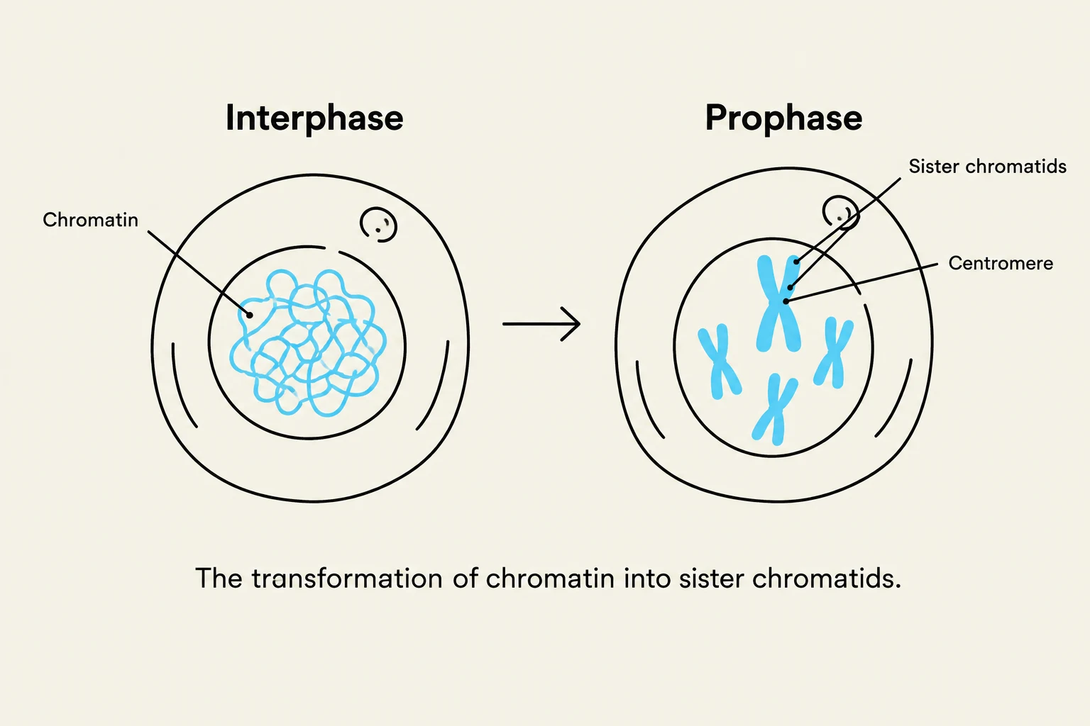
This diagram illustrates the transformation of chromatin into sister chromatids during cell division, showing the stages of Interphase and Prophase.
Before diving into the stages, it is essential to understand the structures involved. During the cell's resting state, DNA exists as a loose, threadlike mass called chromatin. However, as mitosis begins, this chromatin must undergo a transformation to prevent the long DNA strands from entangling during movement.
Through a process of condensation, chromatin fibers shorten into visible chromosomes. Because DNA replication occurs during the S phase of interphase, each chromosome at the start of mitosis consists of two identical strands known as chromatids. These two strands are held together at a constricted region called the centromere.
Study Tip: Terminology Check
It is easy to confuse "chromatid" and "chromosome." Remember: as long as the two identical strands are joined at the centromere, they are called sister chromatids. Once the centromere splits and they move to opposite poles, each individual strand is referred to as a chromosome.
Prophase: Setting the Stage
Prophase is the initial phase where the cellular machinery for division is assembled. Several critical events occur simultaneously:
Condensation: Chromatin fibers condense into visible chromosomes.
Spindle Formation: Tubulins in the centrosome begin to form the mitotic spindle, a football-shaped assembly of microtubules.
Attachment: The spindle microtubules attach to the kinetochore, a protein complex located on the outside of each centromere.
Polarization: As the microtubules lengthen, they push the centromeres toward the opposite poles (ends) of the cell, extending the spindle from pole to pole.
Dissolution: To allow the chromosomes to move freely, the nucleolus disappears and the nuclear envelope breaks down.
Figure 3.32bFigure 3.32b shows the condensation of chromosomes and the formation of the mitotic spindle during prophase.
Metaphase: The Alignment
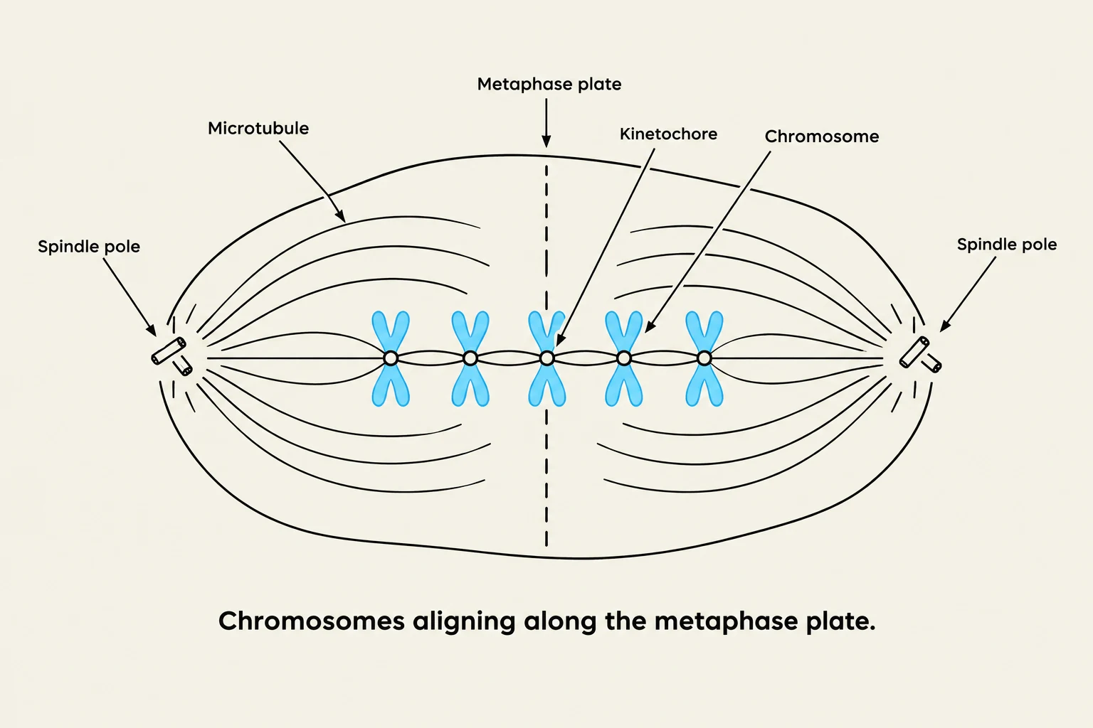
Diagram illustrating chromosomes aligning along the metaphase plate during cell division.
Once the spindle is established, the cell enters metaphase. During this stage, the microtubules of the mitotic spindle work to align the centromeres of the chromatid pairs.
The centromeres are maneuvered to the exact center of the mitotic spindle. This central plane of alignment is known as the metaphase plate (or the equatorial plane). This alignment is a critical checkpoint; if the chromosomes are not properly aligned, the subsequent separation could lead to errors in the amount of DNA each daughter cell receives.
Figure 3.32cFigure 3.32c illustrates the alignment of centromeres along the metaphase plate during metaphase.
Anaphase: The Great Separation
Anaphase is characterized by rapid movement. The centromeres split, which separates the two members of each chromatid pair.
As the mitotic spindle pulls the individual strands toward opposite poles, they are no longer called chromatids—they are now termed chromosomes. A distinctive visual feature of anaphase is that the chromosomes often appear V-shaped. This occurs because the centromeres lead the way, being pulled by the microtubules, while the trailing arms of the chromosomes drag behind.
Figure 3.32dFigure 3.32d shows the separation of chromosomes toward opposite poles during anaphase.
Telophase: Rebuilding the Nucleus
Telophase is the final stage of mitosis, beginning once the chromosomal movement has ceased. This stage essentially reverses many of the processes seen in prophase to return the cell to a stable state:
Decondensation: The identical sets of chromosomes reach the opposite poles, uncoil, and revert to their threadlike chromatin form.
Reconstruction: A new nuclear envelope forms around each mass of chromatin.
Reappearance: The nucleoli reappear within the newly formed, identical nuclei.
Disassembly: The mitotic spindle breaks down.
Figure 3.32eFigure 3.32e depicts the formation of two new nuclei during telophase.
Summary Table of Mitotic Stages
Stage
Key Event
Chromosome State
Prophase
Spindle forms; nuclear envelope breaks down
Condensed into chromatid pairs
Metaphase
Alignment at the metaphase plate
Centromeres at the equator
Anaphase
Centromeres split; chromosomes move to poles
Individual chromosomes (formerly chromatids)
Telophase
Nuclear envelopes reform; spindle breaks down
Uncoiling back into chromatin
Keep reading for free.
Create an account to finish this reading and make it part of your study routine.