The Somatic and Autonomic Nervous Systems: Function and Divisions
An overview of the human nervous system, detailing the functional divisions of the Somatic Nervous System (SNS) and the Autonomic Nervous System (ANS), including the sympathetic and parasympathetic branches. Explains sensory input, motor output, reflexes, homeostasis, and neurotransmitter roles.
The human nervous system is a complex communication network designed to process sensory information and coordinate responses to the environment. To manage the vast array of tasks required for survival—from moving your hand to digesting a meal—the system is functionally divided into two primary branches: the Somatic Nervous System (SNS) and the Autonomic Nervous System (ANS). While one handles our conscious interactions with the world, the other works tirelessly behind the scenes to maintain internal stability, a state known as homeostasis.
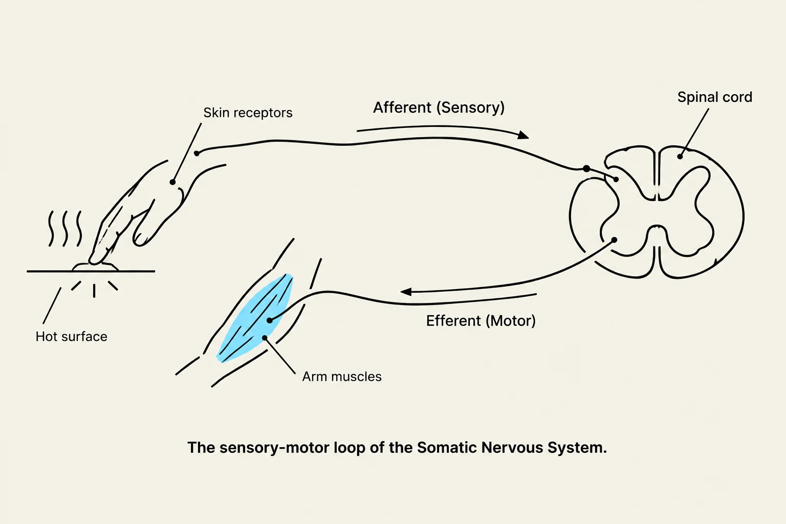
Diagram illustrating the sensory-motor loop of the Somatic Nervous System, showing how skin receptors detect a hot surface, send signals via afferent nerves to the spinal cord, and motor commands are sent back via efferent nerves to arm muscles.
The Somatic Nervous System (SNS)
The Somatic Nervous System is primarily concerned with voluntary control and the transmission of sensory data. It allows us to interact consciously with our surroundings through two main pathways: sensory (afferent) and motor (efferent).
Sensory Input and Motor Output
The SNS utilizes various receptors to gather information about the body and its environment:
Tactile Receptors: These relay information regarding touch, temperature, pain, and pressure.
Proprioceptors: These specialized receptors relay information about the stretch of skeletal muscles, tendons, and joints, helping the brain understand body position.
Once this information reaches the Central Nervous System (CNS), the brain processes it and sends instructions back out via efferent pathways. For example, if you feel a crumb on your lip, mechanoreceptors detect it, sensory nerves (such as the Trigeminal nerve) transmit the signal to the brain, and the brain then coordinates motor nerves (like the Hypoglossal nerve) to move your tongue to remove it.
Reflexes and Plexuses
Not all somatic information requires conscious thought. Reflexes are automatic responses to stimuli that can be either monosynaptic (involving one synapse in the spinal cord) or polysynaptic (involving two or more synapses).
To facilitate communication between the CNS and the Peripheral Nervous System (PNS), the body uses plexuses—networks of nerves that distribute signals to specific regions. Common examples include:
Brachial plexus: Supplies the shoulder, arms, and hands.
Lumbar plexus: Supplies the lower abdominal wall and parts of the lower limbs.
Sacral plexus: Supplies the buttocks, legs, and pelvis.
Exam Tip: Remember the direction of flow! Afferent pathways carry sensory information toward the CNS, while efferent pathways carry motor commands away from the CNS.
The Autonomic Nervous System (ANS)
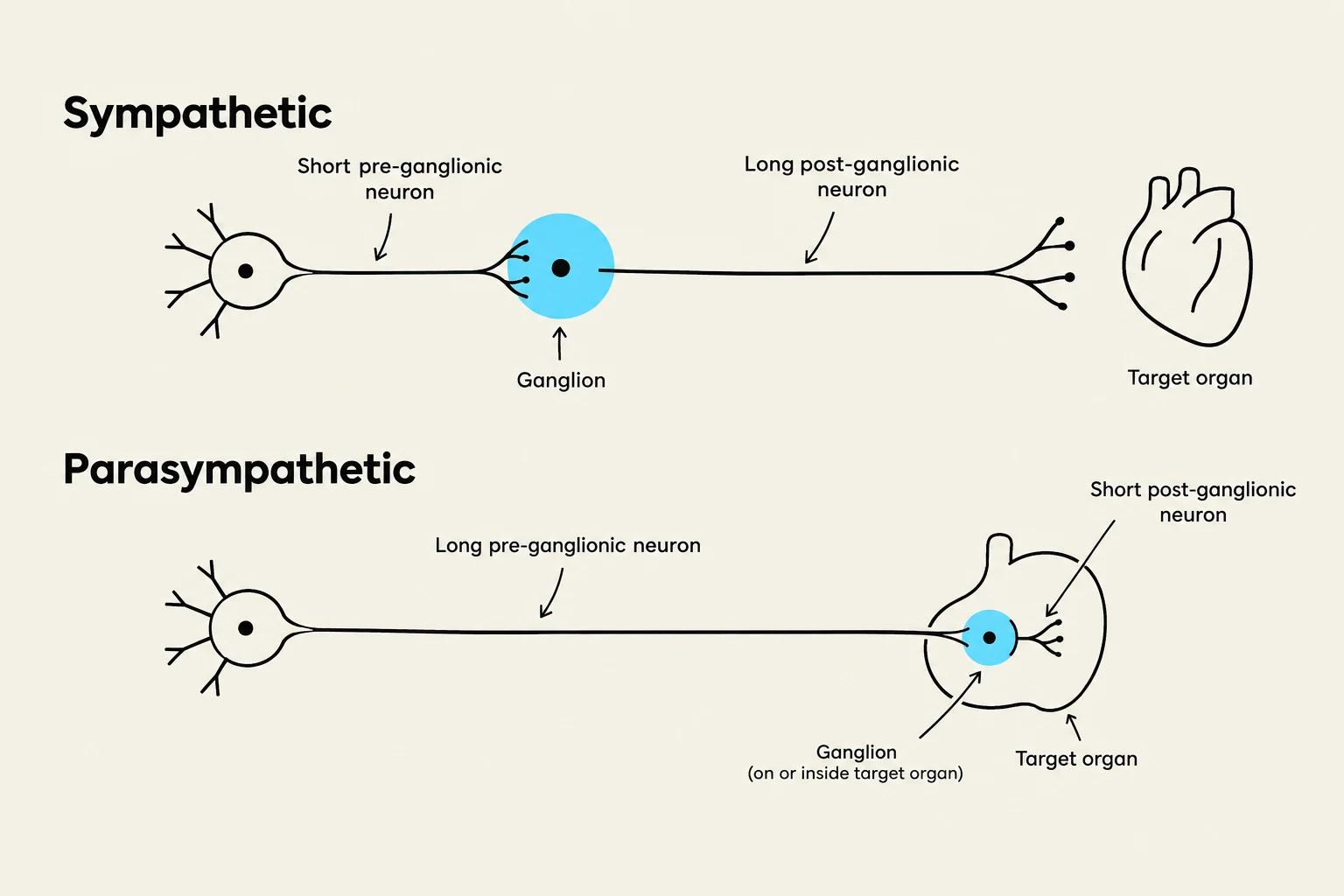
Diagram illustrating the sympathetic and parasympathetic nervous systems, showing the differences in pre-ganglionic and post-ganglionic neuron lengths and ganglion location relative to the target organ.
While the somatic system manages what we do, the Autonomic Nervous System manages what our body does. The ANS governs involuntary physiological functions by regulating smooth muscle, cardiac muscle, and glands. This includes critical processes like heart rate, respiratory rate, and digestion.
The ANS is regulated by the hypothalamus, brainstem, and spinal cord. It is further subdivided into two antagonistic branches that work in opposition to maintain balance: the Sympathetic and Parasympathetic divisions.
The Sympathetic Nervous System: "Fight or Flight"
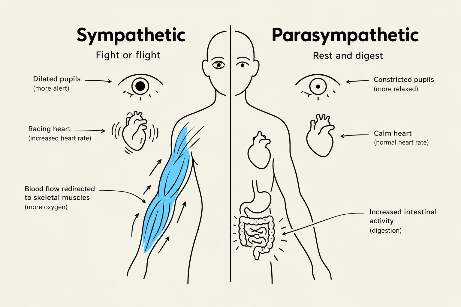
This diagram illustrates the opposing effects of the sympathetic (fight or flight) and parasympathetic (rest and digest) nervous systems on the human body, including pupil dilation/constriction, heart rate, and blood flow redirection.
The Sympathetic Nervous System prepares the body for acute stress or physical activity. When you encounter a perceived threat, this system triggers a systemic response to maximize your ability to survive the encounter. Key physiological changes include:
Increased heart rate.
Dilation of the pupils.
Redirection of blood flow away from the digestive tract and toward the skeletal muscles.
In terms of anatomy, the sympathetic division is characterized by short pre-ganglionic fibers and long post-ganglionic fibers.
The Parasympathetic Nervous System: "Rest and Digest"
Conversely, the Parasympathetic Nervous System is responsible for conserving energy and maintaining "housekeeping" functions. It works to return the body to a state of calm after a stressor has passed. Key functions include:
Slowing the heart rate.
Increasing intestinal activity to promote digestion and waste elimination.
Anatomically, the parasympathetic division features long pre-ganglionic fibers and short post-ganglionic fibers, with the ganglia located very close to or even within the target organs. A major component of this system is the Vagus Nerve (CN X), which innervates most thoracic and abdominal organs.
Chemical Communication: Neurotransmitters
The communication between the autonomic neurons and their target organs relies on specific chemical messengers called neurotransmitters.
Neurotransmitter
Primary Role in the ANS
Acetylcholine (ACh)
Used by the parasympathetic division and at the preganglionic junctions of both divisions.
Norepinephrine (NE)
The primary neurotransmitter used by sympathetic postganglionic neurons to stimulate target organs.
Epinephrine (Epi)
Released from the adrenal medulla into the bloodstream to amplify the systemic sympathetic response.
By utilizing these different chemicals, the body can precisely tune its physiological state—speeding up processes during danger and slowing them down during rest—to ensure the internal environment remains stable.
Keep reading for free.
Create an account to finish this reading and make it part of your study routine.