Explains the sliding filament model of muscle contraction, detailing the roles of sarcomeres, thick and thin filaments (actin, myosin, tropomyosin, troponin), and the molecular mechanisms involving ATP hydrolysis and calcium ions.
Muscle contraction is a highly coordinated mechanical process that allows organisms to move, maintain posture, and circulate blood. At the microscopic level, this movement is not caused by the shortening of individual proteins, but rather by the relative movement of protein filaments sliding past one another. This elegant process, known as the sliding filament model, relies on the precise interaction between thick and thin filaments within the functional units of the muscle fiber.
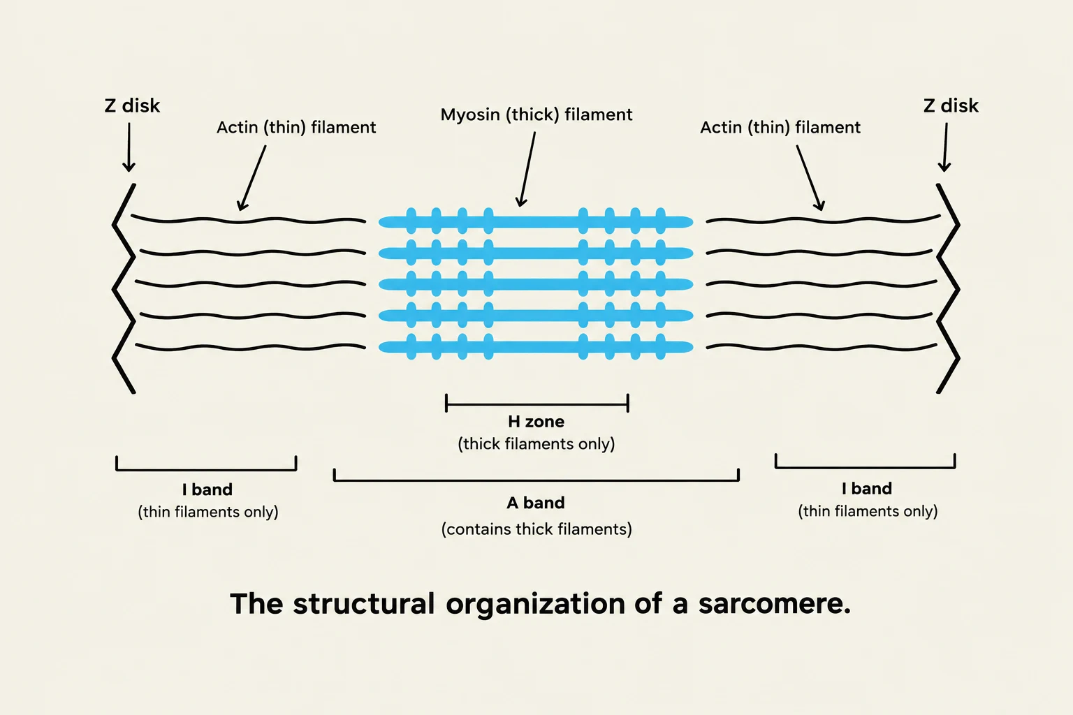
Diagram illustrating the structural organization of a sarcomere, showing the arrangement of actin and myosin filaments, Z disks, H zone, A band, and I band.
The Structural Unit: The Sarcomere
To understand how a muscle contracts, we must first look at the sarcomere, which is the fundamental functional unit of a myofibril. A sarcomere is defined as the segment between two Z disks (or Z lines).
When viewed under an electron microscope, the myofibril displays a distinct pattern of light and dark bands that change during contraction:
A band: This represents the total length of the thick filaments. Importantly, the width of the A band remains constant during contraction.
I band: The light gray areas containing only thin filaments.
H zone: The dark gray area in the center of the A band that contains only thick filaments.
As the muscle contracts, the thin filaments are pulled toward the center of the sarcomere. This causes the I band and the H zone to decrease in width, while the Z disks move closer together, effectively shortening the sarcomere.
The Molecular Machinery: Thick and Thin Filaments
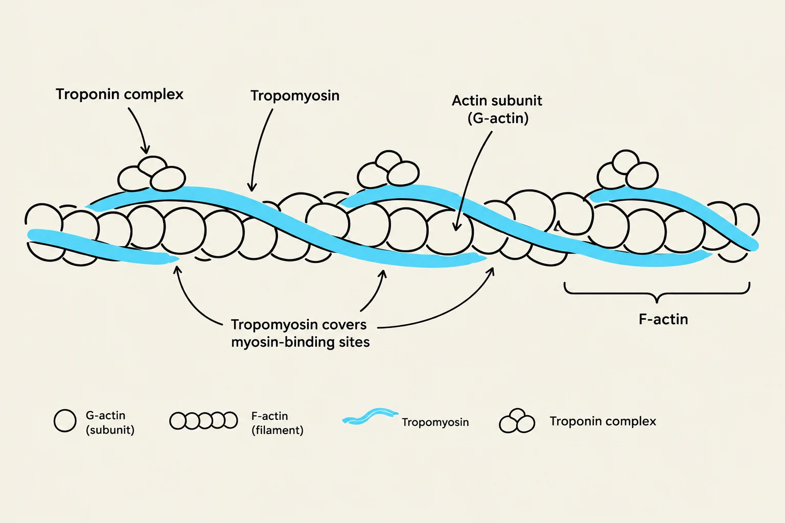
Diagram illustrating the structure of actin filaments, including G-actin subunits, F-actin filaments, tropomyosin, and troponin complex, and how tropomyosin covers myosin-binding sites.
The contraction process is driven by two primary types of protein filaments: the thick filaments and the thin filaments.
Thick Filaments (Myosin)
The thick filament is composed of hundreds of myosin molecules. Each myosin molecule is a complex protein consisting of two heavy chains that form a long, coiled-coil tail (the "backbone" of the filament) and two pairs of light chains.
The most critical part of the myosin molecule for contraction is the myosin head. These heads project outward from the thick filament and serve two vital roles:
They act as ATPases, meaning they are enzymes that catalyze the hydrolysis of ATP to provide energy.
They form cross-bridges by physically binding to the thin filaments.
Thin Filaments (Actin and Regulatory Proteins)
The thin filament is more complex than the thick filament because it contains the regulatory machinery required to "turn on" contraction. It consists of three main components:
Actin: The structural backbone, composed of individual subunits called G-actin that polymerize into a long chain known as F-actin.
Tropomyosin: A coiled-coil protein that wraps around the actin filament. In a relaxed state, tropomyosin covers the specific binding sites where myosin heads would attach.
Troponin: A trimeric protein complex that sits on the tropomyosin. It acts as the "lock" that prevents contraction until the correct signal is received.
Exam Tip: Remember that the filaments themselves do not change length. If a question asks whether actin or myosin shortens during contraction, the answer is no—they simply slide past each other.
The Mechanism of Contraction
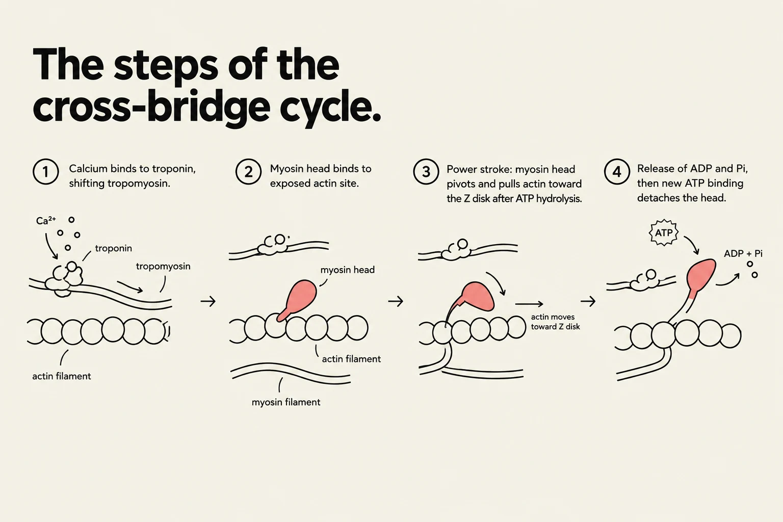
Diagram illustrating the four steps of the cross-bridge cycle in muscle contraction: calcium binding to troponin, myosin head binding to actin, the power stroke, and ATP binding leading to detachment.
Muscle contraction is a cycle of chemical energy conversion into mechanical work, regulated by the presence of calcium ions (Ca2+).
1. The Trigger: Ca2+ Influx
Contraction is initiated when a signal triggers an influx of calcium ions (Ca2+) from the sarcotubules (transverse tubules) into the cytoplasm of the muscle cell.
2. Unblocking the Binding Sites
Once released, the Ca2+ ions bind to troponin. This binding induces a conformational (structural) change in the entire troponin-tropomyosin complex. This change shifts the position of the tropomyosin, moving it away from the myosin-binding sites on the actin filament and exposing them.
3. Cross-bridge Formation and the Power Stroke
With the binding sites exposed, the myosin heads bind to the actin, forming cross-bridges.
The actual "sliding" movement is driven by ATP hydrolysis. The myosin head functions as an enzyme that breaks down ATP into ADP and inorganic phosphate (Pi):
ATP→ADP+Pi
The energy released from this reaction causes the myosin head to undergo a conformational change. This change allows the myosin head to "walk" or pivot along the actin filament toward the Z disk. This movement is known as the power stroke.
4. The Cycle Repeats
As long as Ca2+ is present to keep the binding sites open and ATP is available to provide energy, the myosin heads will continue to attach, pull, release, and reattach, effectively "walking" up the actin filament. This cycle occurs rapidly; approximately 500 heads on every thick filament cycle through this process every second.
Component
Role in Contraction
Myosin
Motor protein; provides force via ATP hydrolysis
Actin
Structural track for myosin to walk upon
Tropomyosin
Inhibitor; blocks myosin binding sites
Troponin
Regulator; binds Ca2+ to move tropomyosin
ATP
Fuel; provides energy for the power stroke
Keep reading for free.
Create an account to finish this reading and make it part of your study routine.