The Skeletal System: Structure, Function, and Repair
An in-depth explanation of the skeletal system, covering its functions, organization into axial and appendicular components, bone structure and composition, cellular activity, growth, remodeling, repair processes, and the role of joints and synovial fluid in movement.
The skeletal system is far more than a rigid scaffolding; it is a dynamic, living organ system essential for survival. From providing the structural framework that maintains body shape to acting as a factory for blood cells, the skeleton integrates mechanical support with complex metabolic functions. Understanding how bones grow, repair, and interact at joints is fundamental to grasping how animals move and maintain homeostasis.
Functions of the Skeleton
The skeleton serves five primary roles that are critical to an organism's physiological stability:
Structural Support: It provides the framework necessary to maintain body shape and offers attachment points for muscles.
Organ Protection: Hard bony structures like the skull, ribcage, and vertebrae shield vital organs such as the brain, heart, lungs, and spinal cord.
Locomotion: The arrangement of bones, particularly in the limbs, is specialized to accommodate different movement styles, such as the digitigrade gait of pigs or the unguligrade gait of cattle.
Mineral Storage: Bones act as a reservoir for essential minerals, specifically calcium and phosphorus, which are vital for metabolic processes.
Hematopoiesis: The bone marrow is the site of blood cell production, a process known as hematopoiesis.
Organization of the Skeleton
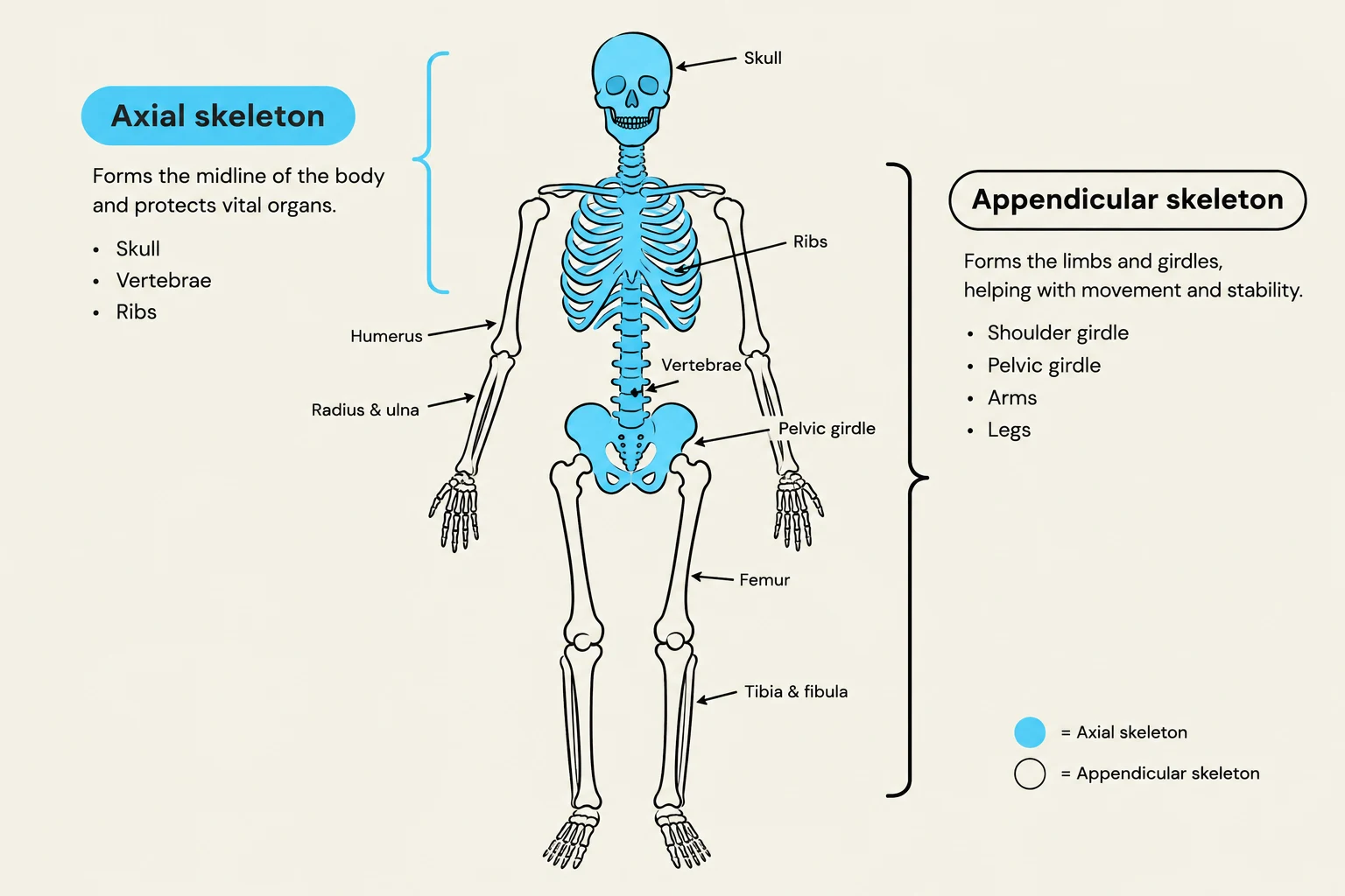
The human and animal skeletons are divided into two main functional groups: the axial and the appendicular skeleton.
The Axial Skeleton
The axial skeleton forms the body's midline or long axis. It includes the skull, the vertebral column, and the bones attached to the vertebrae, such as the ribs and the sternum. Within the vertebral column, the intervertebral discs play a crucial role in spinal health. These discs consist of two parts: the nucleus pulposus, a gel-like center that acts as a shock absorber to distribute pressure, and the anulus fibrosus, a tough outer ring that provides structural containment and resists excessive movement.
The Appendicular Skeleton
The appendicular skeleton comprises the bones of the limbs and the girdles that attach them to the axial skeleton. This includes the pectoral (shoulder) girdle and the pelvic (hip) girdle, which connect the front and hind limbs to the torso.
Diagram illustrating the axial and appendicular skeletons of the human body, labeling key bones and their functions.
Bone Structure and Composition
Bone is a specialized connective tissue with a unique chemical makeup. In terms of wet weight, bone is approximately 25% water, 45% minerals, and 30% organic matter. When looking at dry weight, the mineral component accounts for 65%, with calcium making up about 37% and phosphorus about 18.5%. The organic fraction is largely composed of collagen (about 90%), which provides flexibility.
Anatomical Features
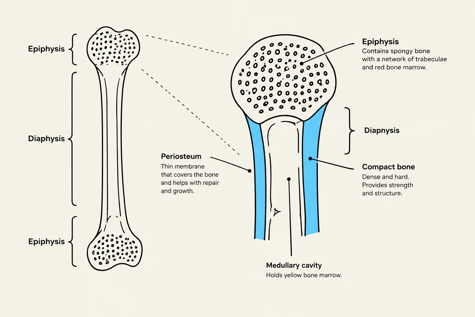
A typical long bone consists of several distinct regions:
Diaphysis: The central shaft of the bone, which provides leverage and strength.
Epiphysis: The expanded ends of the bone that form joints with other bones.
Periosteum: A protective membrane covering the bone surface that supports growth, repair, and nutrient supply.
Marrow Cavity: The central space that may store yellow marrow (fat) or red marrow (blood cell production).
Microscopic Organization: Compact vs. Cancellous Bone
Bone tissue is organized into two main types: compact bone and cancellous (spongy) bone.
Compact bone is dense and provides the primary structural strength. It is organized into functional units called Haversian systems (or osteons):
Haversian Canal: A central channel containing blood vessels, nerves, and lymphatic vessels.
Lamellae: Concentric rings of calcified matrix that surround the canal.
Lacunae: Small spaces located between the lamellae that house osteocytes.
Canaliculi: Tiny, microscopic channels that connect lacunae, allowing osteocytes to exchange nutrients and waste.
Cancellous bone, on the other hand, is a porous, lattice-like structure found primarily in the epiphyses. It houses red marrow for hematopoiesis and provides lightweight support.
Diagram illustrating the structure of a long bone, labeling the epiphysis, diaphysis, periosteum, compact bone, and medullary cavity.
Cellular Activity: The Builders and the Resorbers
Bone is a dynamic tissue that is constantly being created and broken down by four specialized cell types:
Osteoprogenitor Cells: Stem cells that differentiate into osteoblasts.
Osteoblasts: The "builders" that produce and mineralize the bone matrix.
Osteocytes: Mature bone cells that reside in lacunae; they maintain the bone structure and regulate nutrient exchange.
Osteoclasts: The "resorbers" that break down bone tissue to facilitate remodeling and release calcium into the bloodstream.
Exam Tip: Remember the "B" in Osteoblasts stands for "Build." If a question asks which cell is responsible for increasing bone density, the answer is osteoblasts.
Growth, Remodeling, and Repair
Bone Formation and Growth
Long bones grow through a process called ossification. Initially, a model made of hyaline cartilage is formed. Through primary ossification, bone replaces cartilage in the diaphysis. Secondary ossification occurs in the epiphyses after birth. Growth in length continues at the epiphyseal plate (growth plate) until adulthood, when the plate ossifies into the epiphyseal line, signaling that growth has ceased.
Bone Remodeling
Remodeling is the continuous cycle of bone renewal to maintain strength and calcium levels. This cycle follows four stages:
Activation: Osteoclasts are recruited to a specific site.
Resorption: Osteoclasts break down the old or damaged bone.
Reversal: Osteoclasts die off, and osteoblasts arrive at the site.
Formation: Osteoblasts build new bone matrix.
Bone Repair
When a bone fractures, the body undergoes a specific four-step healing process:
Hematoma Formation: A blood clot forms at the fracture site to stabilize the area and bring in repair cells.
Fibrocartilaginous Callus Formation: A "soft callus" made of collagen and cartilage bridges the gap between the broken ends.
Bony Callus Formation: Osteoblasts replace the soft callus with hard, spongy bone.
Bone Remodeling: Osteoclasts reshape the bone to restore its original strength and structure.
Joints and Movement
Joints are the points where bones meet, facilitating movement. To ensure smooth motion, joints are often covered in articular cartilage, which reduces friction and absorbs shock.
The joints are highly innervated to provide feedback to the nervous system. Sensory fibers in the joint capsule and synovial membrane primarily transmit pain signals, while proprioceptive fibers (such as those from Ruffini endings) provide information about the position and movement of the limb.
Diagram illustrating the anatomy of a synovial joint, including bones, articular cartilage, synovial fluid, and the joint capsule.
Keep reading for free.
Create an account to finish this reading and make it part of your study routine.