Mechanotransduction and Potential Mechanosensors
How does a physical weight moving through space become a chemical signal inside a cell? This process is known as mechanotransduction. While scientists have not yet identified every definitive sensor, several proteins are believed to play critical roles in converting mechanical force into hypertrophic signaling:
The Costamere and Dystrophin Complex
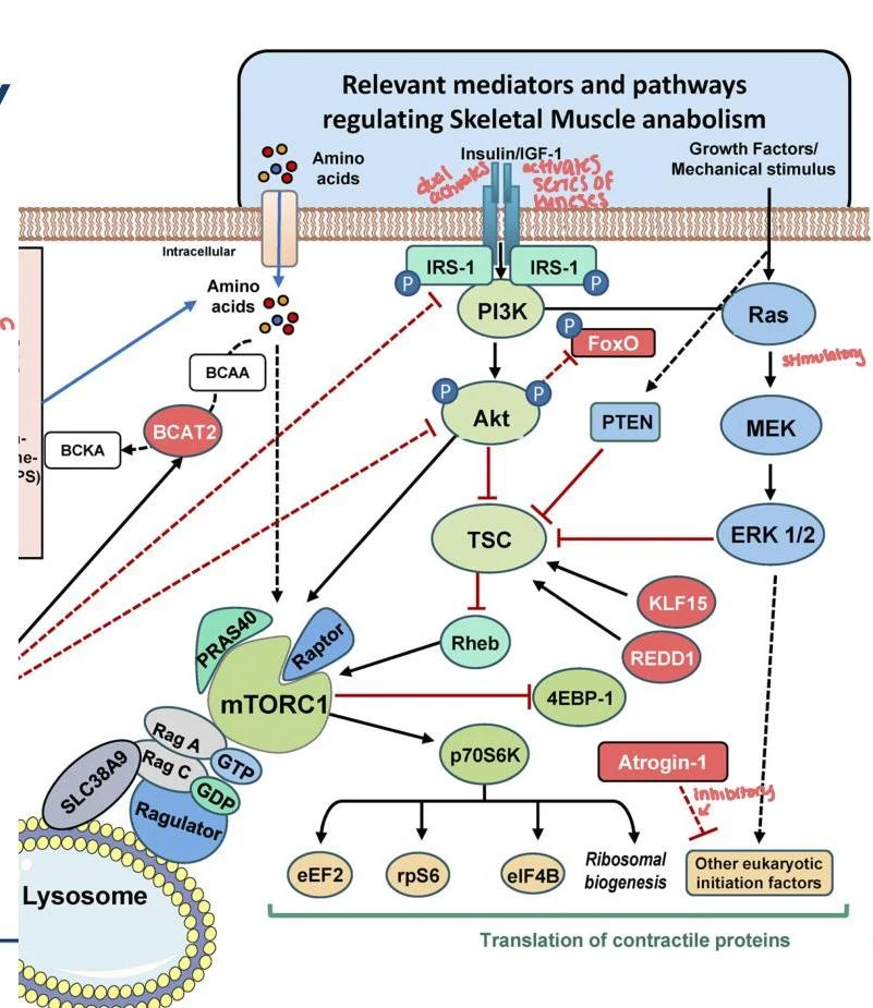
The costamere (involving desmin-ankyrin and integrins) facilitates lateral force transmission across the muscle fiber. Within this structure, the dystrophin protein association complex connects the internal cytoskeleton to the extracellular matrix (ECM). Enzymes such as focal adhesion kinase work at the costamere; they are required for IGF-induced hypertrophy and signaling through the TSC2–mTORC pathway.
Titin
Titin is a massive protein that spans from the Z-disc to the M-line. It acts in parallel with actomyosin force production. While the M-line portion of titin contains a stretch-activated kinase that responds to tension, a definitive link between titin and the activation of mTORC1 has not yet been established.
Filamin-C
Filamin-C attaches actin to the Z-disc. When myosin pulls on actin, Filamin-C deforms, which is postulated to activate mTORC1 via a connection with the protein Bag3 (which inhibits TSC1).
Nuclear Deformation
Mechanical forces can also physically deform the myonuclei. Because myonuclei are anchored to the cytoskeleton, mechanical stress can cause the translocation of signaling proteins into the nucleus through a pathway that is mTORC-independent.
Secondary Stimuli: Damage and Metabolic Stress
While mechanical tension is the primary driver, other factors contribute to the hypertrophic response.
Exercise-Induced Muscle Damage (EIMD)
EIMD is often triggered by novel exercise or eccentric contractions (where the muscle lengthens under load). This can result in microstructural damage, such as Z-disc streaming or membrane damage, which leads to satellite cell activation. However, it is important to note that hypertrophy can occur even in the absence of measurable damage.
Metabolic Stress
The accumulation of metabolites (such as lactate, H+, and Pi) during exercise serves as a potential stimulus. This is the principle behind Blood-Flow Restriction Training (BFRT), where arterial occlusion allows for hypertrophic responses using much lower mechanical loads (e.g., 20% of 1-RM).
Clinical Note: While BFRT is a useful tool for rehabilitation (such as post-ACL reconstruction), research suggests it is generally not superior to traditional strength training (60-80% 1-RM) for muscle hypertrophy.
Practical Application: Dosing Resistance Training
According to the ACSM 2026 Position Stand, the "best" dose depends entirely on your specific goal:
| Goal | Primary Driver | Recommended Dose/Strategy |
|---|
| Muscular Strength | Load/Intensity | Heavier loads (≥80% 1-RM); 2-3 sets; ~2.2 sessions/week |
| Muscle Hypertrophy | Volume | Total weekly volume (≥10 sets per muscle group); wide load range (30-100% 1-RM); include eccentric contractions |
| Muscular Power | Speed/Load | Moderate loads (30-70% 1-RM) with maximal concentric acceleration |
Student Tip: Estimating 1-RM
If you cannot perform a true 1-repetition maximum (1-RM) test, you can estimate it using a repetition maximum (RM) percentage. For example, if you complete 5 repetitions with 100 lbs, and the table indicates 5 reps is 87% of 1-RM, use the following calculation:
0.87100 lbs=1.0x lbs
x=114.94 lbs (Estimated 1-RM)