


Understanding how muscles move is only half the battle in physiology; understanding how they stop moving and how they change in response to stress is equally vital. Muscle function is a delicate balance of chemical signaling, ion concentration, and metabolic availability. This article explores the systematic process of muscle relaxation, the biological phenomenon of rigor mortis, and the various ways muscle tissue adapts to different types of physical stimuli.
The Physiology of Muscle Relaxation
Muscle relaxation is not merely the absence of contraction; it is an active, systematic physiological process that reverses the events of contraction. To stop a contraction, the body must terminate the electrical signal, reset the membrane potential, and remove the chemical triggers from the contractile machinery.
Termination of the Signal
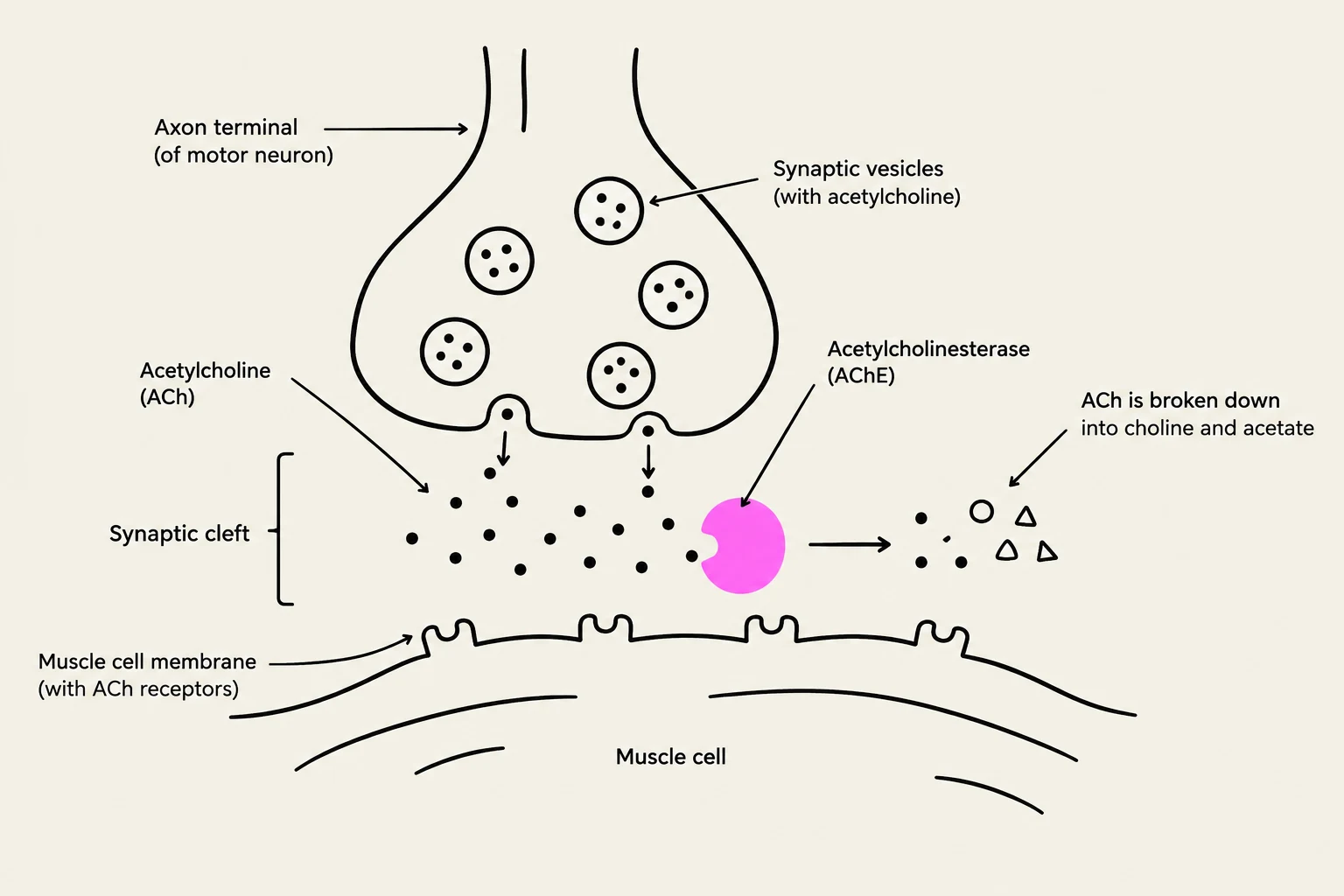
The process begins at the neuromuscular junction (NMJ). For a muscle to relax, the release of the neurotransmitter acetylcholine (ACh) from the motor neuron must cease. However, even after the nerve stops firing, ACh remains in the synaptic cleft, which could potentially continue to stimulate the muscle. To prevent this, the enzyme acetylcholinesterase (AChE) rapidly degrades the remaining ACh.
Once the ACh is cleared, the sarcolemma (the muscle cell membrane) undergoes repolarization. This means the membrane potential returns to its resting state, effectively "resetting" the electrical environment of the muscle fiber.
Calcium Sequestration and Crossbridge Inhibition
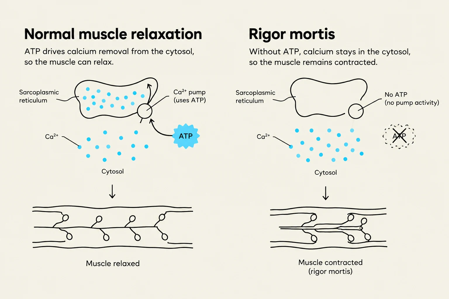
The most critical step in relaxation involves the management of calcium ions (). During contraction, calcium is released into the sarcoplasm to trigger movement. To relax, this calcium must be removed. This is achieved by SERCA pumps (Sarco/Endoplasmic Reticulum -ATPase), which use active transport to pump back into the sarcoplasmic reticulum (SR).

Keep reading for free.
Create an account to finish this reading and make it part of your study routine.
Continue reading freeAlready have an account? Log in
Updated