The Nerve-Muscle Relationship and Muscle Contraction
Explores the intricate communication between motor neurons and muscle fibers, the electrochemical principles of cell stimulation, and the mechanical phases of muscle contraction, including the neuromuscular junction, action potentials, and the effects of toxins.
Skeletal muscle is a highly specialized tissue designed for movement, but it cannot function in isolation. For a muscle to contract, it must receive a specific electrical signal from the nervous system. This article explores the intricate communication between motor neurons and muscle fibers, the electrochemical principles that allow cells to respond to stimuli, and the mechanical phases that turn an electrical impulse into physical force.
The Motor Unit: The Functional Building Block
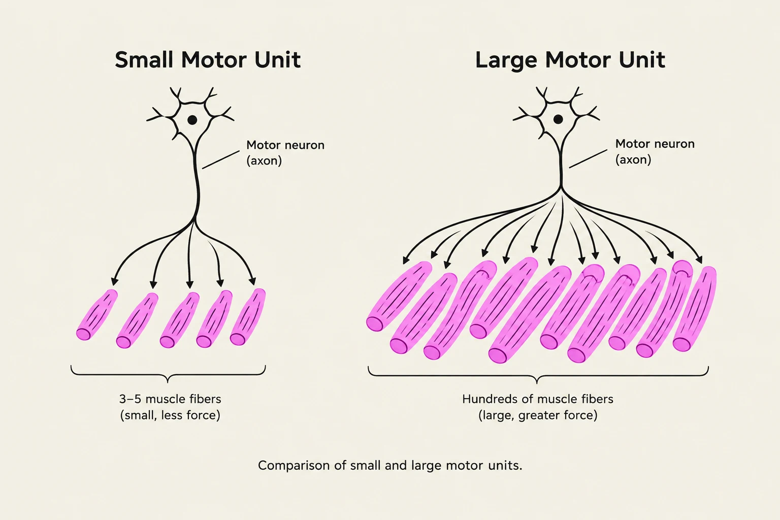
Comparison of small and large motor units, illustrating the difference in the number of muscle fibers innervated by a single motor neuron.
Skeletal muscles are controlled by somatic motor neurons, which are nerve cells with cell bodies located in the brainstem or spinal cord. These neurons send axons, known as somatic motor fibers, directly to the skeletal muscles.
A single nerve fiber does not act alone; instead, it branches out to innervate several muscle fibers. The combination of one nerve fiber and all the muscle fibers it innervates is called a motor unit. All fibers within a single motor unit contract in unison, acting as a functional unit.
The size of a motor unit determines the type of movement possible:
Small Motor Units: These consist of only a few muscle fibers (e.g., 3 to 5) and are found in muscles requiring fine, precise control, such as those in the eyes or hands.
Large Motor Units: These contain hundreds or even thousands of muscle fibers (e.g., the quadriceps) and are designed for generating great strength rather than precision.
Clinical Note: Denervation Atrophy
If the connection between a nerve and a muscle is severed or lost, the muscle loses its stimulation. This leads to denervation atrophy, a process where the paralyzed muscle begins to shrink and waste away.
The Neuromuscular Junction (NMJ)
Diagram illustrating the structure and chemical signaling at the neuromuscular junction, showing the axon terminal, synaptic cleft, and sarcolemma with neurotransmitter release and receptor binding.
The point where the somatic motor neuron meets the muscle fiber is a specialized synapse called the neuromuscular junction (NMJ), also known as the motor end plate. This junction is composed of three critical structural components:
Axon Terminal: The swollen end of the nerve fiber that contains synaptic vesicles filled with the neurotransmitter acetylcholine (ACh).
Synaptic Cleft: The narrow, fluid-filled gap between the axon terminal and the muscle fiber.
Sarcolemma: The plasma membrane of the muscle fiber. To maximize the efficiency of communication, the sarcolemma features junctional folds that increase the surface area, allowing for a higher density of ACh receptors.
When a nerve impulse reaches the axon terminal, ACh is released via exocytosis into the synaptic cleft. The ACh then binds to receptors on the sarcolemma to trigger a response. To ensure that the muscle does not stay in a state of constant contraction, an enzyme called acetylcholinesterase (AChE) resides in the cleft to rapidly degrade ACh, thereby terminating the signal and allowing for relaxation.
Electrical Excitability and the Action Potential
Both nerve and muscle cells are electrically excitable, meaning they can change their membrane voltage in response to chemical or electrical stimuli. This excitability is driven by differences in ion concentrations between the intracellular fluid (ICF) and the extracellular fluid (ECF).
In a resting state, the cell is polarized. It maintains a resting membrane potential (RMP), which is a net negative charge inside the cell compared to the outside. This state is maintained by a high concentration of potassium ions ($K^+$) inside the cell and a high concentration of sodium ions (Na+) outside the cell.
When the NMJ is stimulated, the following electrical sequence occurs:
Depolarization: Ion channels open, allowing Na+ to rush into the cell down its electrochemical gradient, making the inside of the membrane less negative.
Repolarization: Additional channels allow $K^+$ to flow out of the cell, restoring the negative internal charge.
Action Potential: This rapid up-and-down shift in voltage is called an action potential, a wave of electrical excitation that travels along the muscle fiber.
The Four Phases of Muscle Activity
Diagram illustrating the four phases of muscle contraction: excitation, excitation-contraction coupling, contraction, and relaxation, detailing the roles of the sarcolemma, sarcoplasmic reticulum, calcium ions, and actin and myosin filaments.
The transition from an electrical signal to a mechanical contraction follows a highly regulated four-phase process:
Excitation: The action potential travels along the motor nerve and subsequently along the muscle fiber's sarcolemma.
Excitation–Contraction Coupling: This is the vital link where the action potential on the sarcolemma triggers the release of calcium from the sarcoplasmic reticulum (SR), which in turn activates the myofilaments (actin and myosin).
Contraction: The myofilaments slide past one another, causing the muscle fiber to develop tension and potentially shorten.
Relaxation: Once the stimulation ends and ACh is degraded, calcium is pumped back into the SR, and the muscle returns to its resting length.
Neuromuscular Toxins and Paralysis
Because the NMJ relies on a precise balance of neurotransmitters and receptors, certain toxins can disrupt this process, leading to paralysis.
Spastic Paralysis
Spastic paralysis is a state of continual, uncontrolled muscle contraction. This can be caused by toxins like tetanus (Clostridium tetani). The tetanus toxin blocks the release of glycine in the spinal cord; since glycine is an inhibitory neurotransmitter that normally stops motor neurons from firing, its absence leads to overstimulation and constant contraction.
Flaccid Paralysis
Flaccid paralysis is a state where muscles are limp and cannot contract. This can occur through two different mechanisms:
Receptor Blockage: The toxin curare competes with ACh for the receptor sites on the sarcolemma. It binds to the receptor but does not trigger a response, effectively "plugging" the site so ACh cannot work.
Release Blockage:Botulism (caused by Clostridium botulinum) prevents the release of ACh from the axon terminal entirely, meaning no signal ever reaches the muscle.
Summary Table: Types of Paralysis
Type
Muscle State
Mechanism Example
Spastic
Continual contraction
Tetanus (blocks inhibitory glycine)
Flaccid
Limp, no contraction
Curare (blocks ACh receptors) or Botulism (blocks ACh release)
Keep reading for free.
Create an account to finish this reading and make it part of your study routine.