The Muscular System: Structure, Function, and Contraction
An overview of the muscular system, detailing the three types of muscle tissue, the hierarchical structure of skeletal muscle, the mechanism of muscle contraction via the sliding filament theory, metabolic energy production, and adaptations like hypertrophy and atrophy.
The muscular system is a complex network of tissues responsible for generating force, producing movement, and maintaining posture. While we often think of muscles simply as the "strength" behind our actions, they are sophisticated organs composed of muscle tissue, nervous tissue, blood, and connective tissue. To understand how we move, we must distinguish between the three distinct types of muscle tissue: skeletal (voluntary and striated), cardiac (involuntary and striated), and smooth (involuntary and nonstriated).
The Three Types of Muscle Tissue
Each muscle type is specialized for its specific role in the body:
Skeletal Muscle: These are voluntary muscles attached to bones. They are characterized by a striated (banded) appearance and are composed of long, fused cells known as muscle fibers or myofibers.
Cardiac Muscle: Found exclusively in the heart, these cells (called cardiomyocytes) are striated and involuntary. They are arranged in branching, three-dimensional networks and are self-exciting and rhythmic.
Smooth Muscle: These are involuntary and lack striations, giving them a "smooth" appearance. The cells are fusiform (tapered at the ends). They are categorized into multiunit (e.g., blood vessels, iris) and visceral (e.g., stomach, intestines). Visceral smooth muscle often exhibits peristalsis, a wavelike motion that moves contents through tubular organs.
Hierarchical Structure of Skeletal Muscle
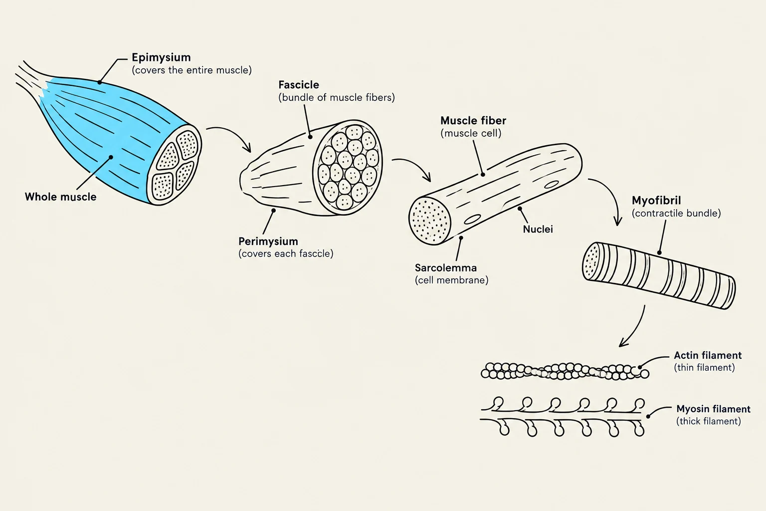
Skeletal muscle is organized into a strict hierarchy, protected and separated by layers of dense connective tissue called fascia. This tissue can form cord-like tendons that attach to the bone's periosteum, or broad sheets called aponeuroses that connect muscles to one another.
The internal organization follows a specific pattern of nesting:
Epimysium: The outermost layer surrounding the entire muscle.
Perimysium: An internal layer that bundles muscle fibers into groups called fascicles.
Endomysium: The innermost layer covering each individual muscle fiber.
Within each muscle fiber, the structure continues down to the molecular level. Each fiber contains 4–20 rod-like filaments called myofibrils. These myofibrils are composed of repeating functional units called sarcomeres, which create the characteristic light and dark "striated" pattern.
Diagram illustrating the hierarchical structure of a muscle, from the whole muscle down to the actin and myosin filaments within a myofibril.
The Sarcomere: The Functional Unit
The sarcomere is the segment between two Z lines (or Z discs). It consists of two main types of protein filaments:
Thick Filaments: Composed of the protein myosin.
Thin Filaments: Composed primarily of the protein actin, along with regulatory proteins troponin and tropomyosin.
The arrangement creates I bands (light bands containing only thin actin filaments) and A bands (dark bands containing thick myosin filaments that overlap with actin).
Muscle Roles and Movement
Muscles rarely work in isolation; they function in groups to produce smooth, controlled movement. To understand these interactions, we look at the relationship between the origin (the fixed, usually proximal attachment) and the insertion (the movable, usually distal attachment).
When a muscle contracts, it pulls the insertion toward the origin. Within a functional group, muscles take on specific roles:
Agonist (Prime Mover): The muscle providing the primary force for a specific movement.
Synergist: An assistant muscle that stabilizes joints or helps the prime mover.
Antagonist: A muscle that opposes the prime mover. For smooth motion, the antagonist must relax while the agonist contracts.
Exam Tip: Remember that muscle roles are relative to the action. A muscle can be an agonist for one movement (e.g., elbow flexion) and an antagonist for another (e.g., elbow extension).
The Mechanism of Contraction
The Neuromuscular Junction
Contraction begins when a motor neuron stimulates a muscle fiber at the neuromuscular junction. This synapse involves a specialized region of the muscle membrane called the motor end plate.
When a nerve impulse reaches the end of a motor neuron, it triggers the release of the neurotransmitter acetylcholine (ACh) into the synaptic cleft. ACh binds to receptors on the motor end plate, causing sodium to flow into the cell, which results in depolarization. This electrical impulse (action potential) travels across the sarcolemma (cell membrane) and deep into the fiber through transverse (T) tubules.
The Sliding Filament Theory
The actual shortening of the muscle is explained by the sliding filament theory.
Calcium Release: The action potential traveling through the T-tubules triggers the sarcoplasmic reticulum to release stored calcium ions into the sarcoplasm.
Regulatory Shift: Calcium binds to troponin, causing a shape change that moves tropomyosin away from the binding sites on the actin filament.
Cross-Bridge Formation: With the binding sites exposed, the myosin heads attach to the actin, forming "cross-bridges".
The Power Stroke: Using energy from ATP, the myosin heads pivot, pulling the actin filaments toward the center of the sarcomere (the M line).
Detachment: A new molecule of ATP binds to the myosin head, causing it to release the actin so the cycle can repeat.
Metabolic Energy for Contraction
Muscle contraction is an energy-intensive process requiring ATP. Because muscle fibers only store enough ATP for 1–2 seconds of activity, the body must constantly regenerate it through three main pathways:
Creatine Phosphate: For immediate, short-term energy (lasting roughly 3–15 seconds), the enzyme creatine kinase transfers a phosphate from creatine phosphate to ADP to rapidly create ATP.
Anaerobic Respiration (Glycolysis): When oxygen is limited, the cell breaks down glucose without oxygen. This produces ATP quickly but results in the accumulation of lactate.
Aerobic Respiration: For sustained, long-term activity, the cell utilizes oxygen in the mitochondria through the Krebs cycle and the Electron Transport System. This is the most efficient way to produce ATP.
Adaptation and Fatigue
Hypertrophy vs. Atrophy
Muscles adapt to the demands placed upon them:
Hypertrophy: An increase in muscle size and strength resulting from increased activity (specifically, an increase in the size of existing fibers and the number of actin/myosin filaments).
Atrophy: A decrease in muscle size and strength due to disuse or inactivity.
Fatigue and Soreness
Muscle fatigue is a transient loss of work capacity where the muscle can no longer contract forcefully. This can be caused by depletion (running out of ATP, CP, or glycogen) or accumulation (the buildup of hydrogen ions, ammonia, or phosphate). Intense exertion can also lead to oxygen debt, where the body must consume extra oxygen after exercise to metabolize accumulated lactate.
Muscle soreness is categorized into two types:
Acute Soreness: Pain during or immediately after exercise, often caused by metabolic by-products.
Delayed-Onset Muscle Soreness (DOMS): Pain that peaks 24–48 hours after exercise. Theories suggest DOMS is caused by mechanical trauma to the muscle fibers or local ischemia (reduced blood flow) due to swelling.
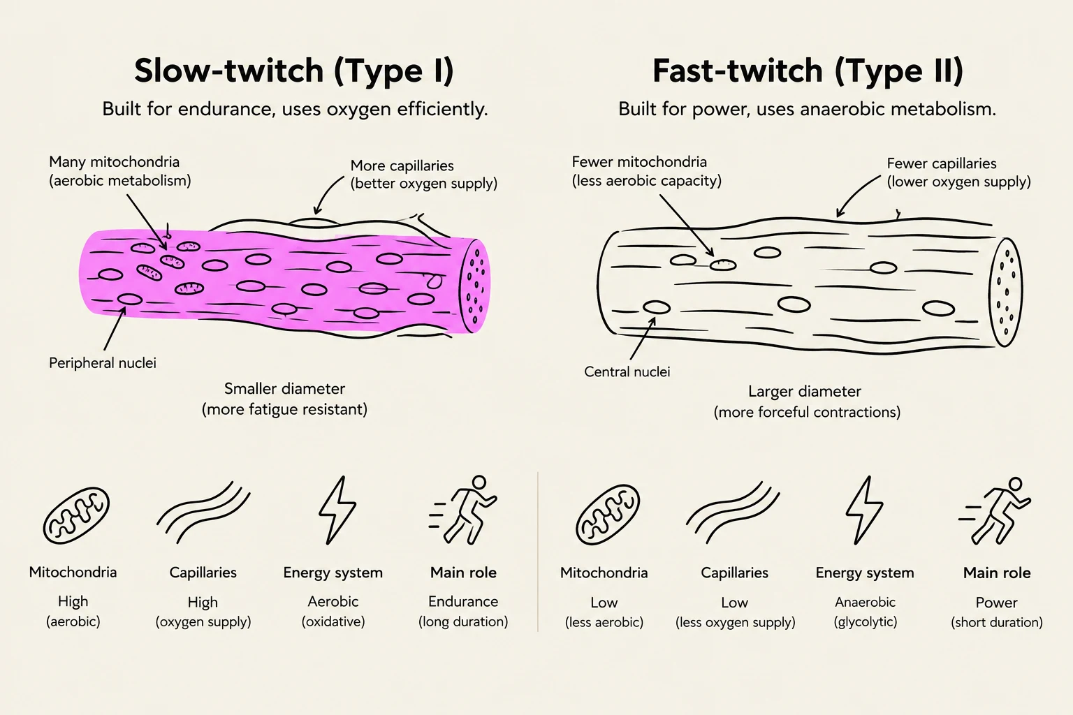
This illustration compares slow-twitch (Type I) and fast-twitch (Type II) muscle fibers, detailing their structural differences, energy systems, and primary roles in the body.
Summary Table: Muscle Comparison
Feature
Skeletal
Smooth
Cardiac
Control
Voluntary
Involuntary
Involuntary
Appearance
Striated
Nonstriated
Striated
Cell Shape
Long, cylindrical
Fusiform (tapered)
Branching networks
Nuclei
Many
Single
Single
Keep reading for free.
Create an account to finish this reading and make it part of your study routine.