The process of emptying the bladder, known as urination or voiding, is not merely a passive event. It is a highly coordinated physiological process governed by the nervous system to ensure that urine is expelled efficiently and at appropriate times. This coordination is achieved through the micturition reflex, a complex interplay between involuntary smooth muscle contractions and voluntary skeletal muscle control.
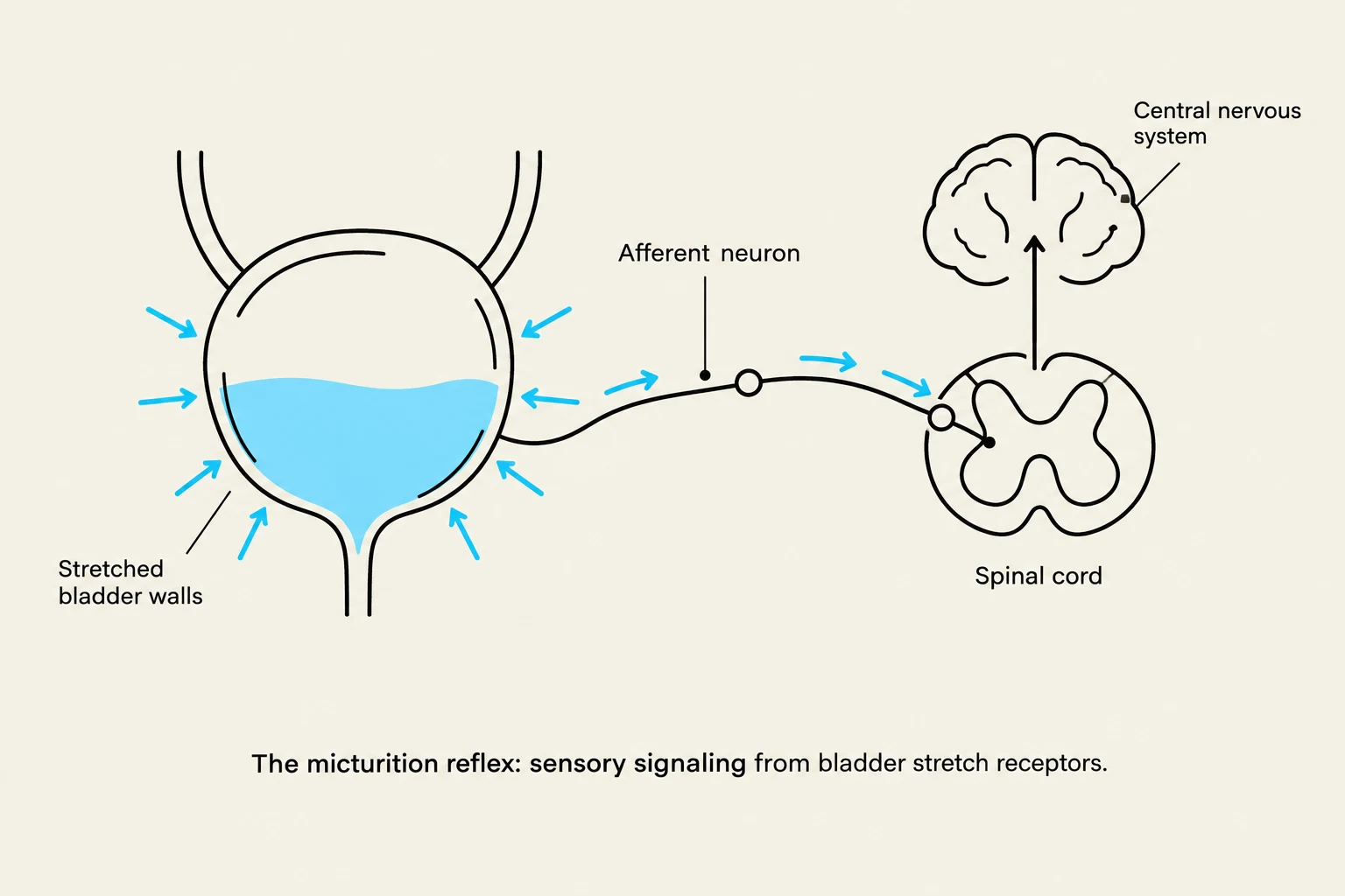
The micturition reflex: sensory signaling from bladder stretch receptors.
Understanding this reflex requires a clear distinction between the different muscle layers of the bladder and the specific neural pathways that command them.
The Micturition Reflex: An Overview
The micturition reflex is a physiological process governed by the autonomic nervous system that facilitates the emptying of the urinary bladder. At its core, the reflex is designed to respond to the stretching of the bladder wall as it fills with urine. Once a certain threshold of fullness is reached, sensory signals are sent to the spinal cord, triggering a coordinated response to initiate voiding.
This reflex is primarily mediated by parasympathetic pathways. The parasympathetic nervous system is responsible for "rest and digest" functions, which in the context of the urinary system, translates to the "emptying" phase.
The Core Mechanism: Two Opposing Actions
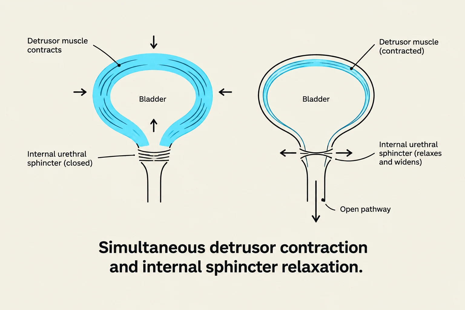
Diagram illustrating the simultaneous contraction of the detrusor muscle and relaxation of the internal urethral sphincter, leading to an open pathway for urination.
For successful urination to occur, the body must perform two simultaneous and opposing actions: it must create pressure within the bladder while simultaneously opening the exit path. This is achieved through the following mechanical changes:
1. Contraction of the Detrusor Muscle
The detrusor muscle is the smooth muscle layer that forms the wall of the urinary bladder. During the micturition reflex, parasympathetic stimulation causes the detrusor muscle to contract. As this muscle layer tightens, it increases the intravesical pressure (the pressure inside the bladder), which is necessary to force urine out of the organ.
2. Relaxation of the Internal Urethral Sphincter
While the bladder wall is contracting, the exit must be opened. The internal urethral sphincter is an involuntary smooth muscle located at the junction of the bladder and the urethra. As part of the parasympathetic reflex, this sphincter relaxes, removing the resistance to urine flow and allowing the liquid to move from the bladder into the urethra.
Exam Tip: The "Result" Question
On many anatomy and physiology exams, you may be asked: "What is the result of the micturition reflex?"
Always look for the combination of detrusor contraction AND internal sphincter relaxation. A common distractor is to suggest the relaxation of the external sphincter; while that happens during voluntary urination, it is not the defining physiological result of the involuntary reflex itself.
Distinguishing the Sphincters
Diagram comparing the internal and external urethral sphincters, illustrating their location and muscle type (involuntary smooth muscle for internal, voluntary skeletal muscle for external).
One of the most critical concepts in studying the micturition reflex is distinguishing between the two different sphincters that control the flow of urine. They differ in tissue type, control mechanism, and their role in the reflex.
Feature
Internal Urethral Sphincter
External Urethral Sphincter
Muscle Type
Smooth Muscle
Skeletal Muscle
Control Type
Involuntary (Autonomic)
Voluntary (Somatic)
Role in Reflex
An effector of the parasympathetic reflex
Not an effector of the reflex; used for conscious control
The Internal Urethral Sphincter
Because it is composed of smooth muscle, the internal sphincter is under the direct command of the autonomic nervous system. It acts as an effector of the micturition reflex, meaning it automatically responds to the parasympathetic signals to relax when the reflex is triggered.
The External Urethral Sphincter
The external sphincter is composed of skeletal muscle, which means it is under voluntary control via the somatic nervous system. While this muscle plays a vital role in our ability to delay urination (by consciously squeezing the muscle to hold urine), it is not considered an effector of the involuntary micturition reflex itself.
Summary of the Parasympathetic Reflex Pathway
To visualize the process, follow this sequence:
Bladder Filling: Urine enters the bladder, stretching the detrusor muscle.
Sensory Input: Stretch receptors send signals to the central nervous system.
Parasympathetic Activation: The parasympathetic nervous system sends motor signals back to the bladder.
Effector Response:
The detrusor muscle contracts (increasing pressure).
The internal urethral sphincter relaxes (opening the path).
Voluntary Check: The brain decides whether to relax the external urethral sphincter to allow the urine to exit the body.
Keep reading for free.
Create an account to finish this reading and make it part of your study routine.