The Lymphoid and Immune Systems: Fluid Recovery and Defense
An overview of the human lymphoid and immune systems, detailing their roles in fluid recovery, defense against pathogens, and absorption of dietary lipids. Covers lymphatic vessels, organs, and the three lines of innate and adaptive immunity.
The human body is constantly exposed to a variety of pathogens, including viruses, bacteria, and fungi. To survive, we rely on a sophisticated, multi-layered defense network known as the immune system. Working in tandem with this defense is the lymphoid system, a complex network of vessels, tissues, and organs that facilitates fluid recovery, immune surveillance, and the absorption of dietary lipids. Together, these systems ensure that the body maintains fluid homeostasis while actively identifying and neutralizing threats.
The Lymphoid System: Vessels and Fluid Flow
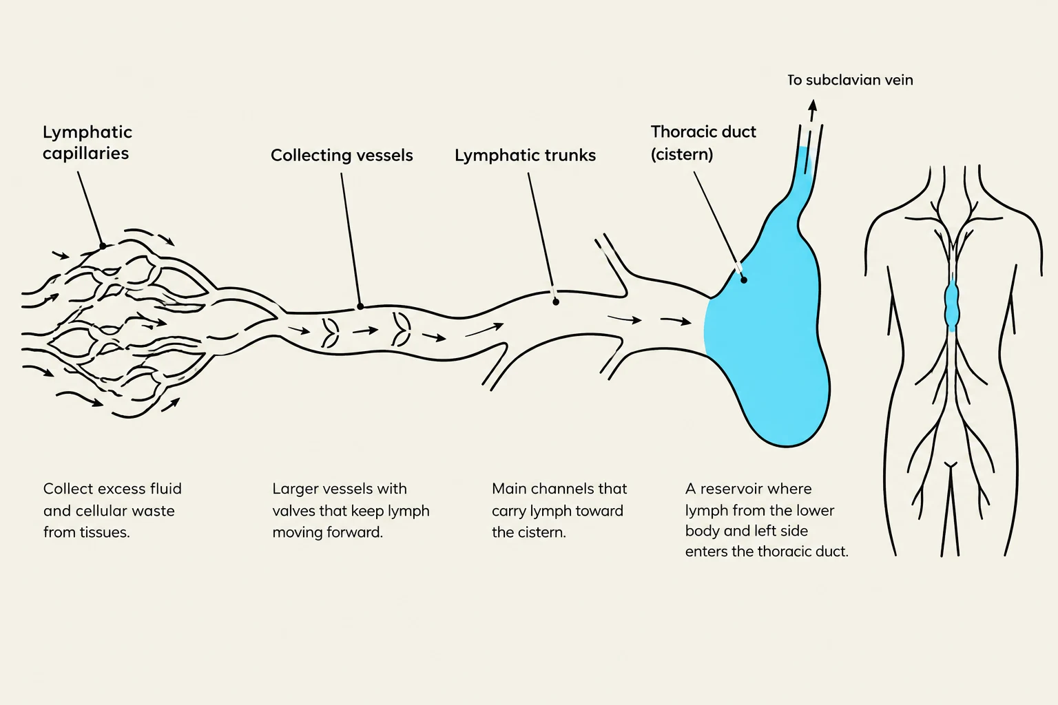
The lymphatic system begins at the microscopic level with lymphatic capillaries. These are highly permeable vessels that penetrate nearly every tissue in the body. Their primary role is to collect excess interstitial fluid—the fluid that filters out of blood capillaries into tissue spaces—and turn it into lymph. Lymph is a clear, colorless fluid that is similar to plasma but contains significantly lower levels of protein. In the small intestine, specialized lymphatic capillaries called lacteals play a critical role in absorbing dietary lipids.
Once collected, lymph moves from these capillaries into larger collecting vessels, which eventually merge into lymphatic trunks. These trunks drain major portions of the body and converge into two large collecting ducts:
The Right Lymphatic Duct: Receives lymph from the right arm, right side of the head, and the right thorax, emptying into the right subclavian vein.
The Thoracic Duct: The larger and longer of the two, it begins as a sac in the abdomen called the cisterna chyli. It receives lymph from below the diaphragm, the left arm, and the left side of the head, neck, and thorax, eventually emptying into the left subclavian vein.
Diagram illustrating the lymphatic system, showing lymphatic capillaries, collecting vessels, lymphatic trunks, and the thoracic duct leading to the subclavian vein.
Because the lymphatic system lacks a central pump like the heart, lymph flows at a lower pressure and slower speed than venous blood. Movement is driven by the rhythmic contractions of lymphatic vessels, the squeezing of skeletal muscles, the pulsations of nearby arteries, and the "thoracic pump" created by breathing.
Exam Tip: Remember that while blood capillaries reabsorb about 85% of filtered fluid, the remaining 15% must be recovered by the lymphatic system to prevent tissue swelling (edema).
In the brain, a specialized lymphatic-resembling drainage pathway known as the glymphatic system exists, where lymphatic-resembling vessels associated with neuroglia help manage fluid within the central nervous system.
Lymphoid Organs and Tissues
Lymphoid structures are categorized based on their function in the immune response.
Primary Lymphoid Organs
These are the sites where immune cells are produced and undergo maturation to become "immunocompetent" (capable of recognizing specific antigens).
Red Bone Marrow: The site of hematopoiesis (blood cell formation), where all lymphocytes originate. B cells also complete their development here.
Thymus: A bilobed organ located in the superior mediastinum. It is the site where T cells mature and undergo rigorous testing (positive and negative selection) to ensure they can recognize foreign antigens without attacking the body's own tissues.
Secondary Lymphoid Organs
These are the "battlegrounds" where mature immune cells encounter pathogens and become activated.
Lymph Nodes: Bean-shaped structures that cleanse lymph as it passes through them. They are major sites for lymphocyte activation.
Tonsils: Patches of lymphoid tissue at the entrance to the pharynx that guard against inhaled or ingested pathogens. They contain deep pits called tonsillar crypts that increase surface area for trapping microbes.
Spleen: The body's largest lymphoid organ. It contains red pulp, which acts as an "erythrocyte graveyard" by filtering out old or fragile red blood cells, and white pulp, which monitors the blood for foreign antigens.
Beyond these organized organs, lymphoid tissue can be diffuse, such as mucosa-associated lymphoid tissue (MALT), which is found in the mucous membranes of the respiratory, digestive, urinary, and reproductive tracts.
The Three Lines of Defense
The immune system operates through three distinct lines of defense to protect against pathogens.
1. The First Line: Innate Barriers
This is the immediate, nonspecific defense consisting of physical and chemical barriers, primarily the skin (due to its tough keratin) and mucous membranes.
2. The Second Line: Innate Immunity
If a pathogen breaches the physical barriers, the second line of defense is activated. This is a nonspecific response that includes:
Leukocytes (White Blood Cells): Including phagocytes (like neutrophils and macrophages) that engulf foreign matter, and natural killer (NK) cells that destroy infected or cancerous host cells.
Antimicrobial Proteins: Such as the complement system (a group of 30+ proteins that assist in inflammation and cell destruction) and interferons (proteins secreted by virally infected cells to warn neighboring cells).
Inflammation and Fever: Inflammation is a local response to tissue injury characterized by heat, redness, swelling, and pain. Fever (pyrexia) is an abnormal elevation in body temperature that can help inhibit pathogen growth.
3. The Third Line: Adaptive Immunity
Adaptive immunity is highly specific and provides long-lasting protection through immunological memory. It is characterized by its ability to distinguish "self" from "nonself" and its systemic effect throughout the body. It is divided into two branches:
Cellular Immunity
Mediated by T lymphocytes (T cells), this branch involves cells that directly attack and destroy diseased or foreign cells. This process relies on antigen-presenting cells (APCs), such as macrophages or dendritic cells, which use major histocompatibility complex (MHC) proteins to "show" antigens to T cells.
Humoral Immunity
Mediated by B lymphocytes (B cells) and the antibodies (immunoglobulins) they produce. When a B cell is activated, it differentiates into plasma cells, which secrete massive amounts of antibodies into the blood and lymph. These antibodies can neutralize toxins, fix complement, or tag pathogens for destruction.
Key Concept: Memory
One of the most critical aspects of adaptive immunity is the creation of memory cells. During a primary immune response (the first exposure to a pathogen), the body takes time to build up an antibody titer. However, during a secondary (anamnestic) response (re-exposure), memory cells allow the body to react so rapidly that the individual often does not even realize they were exposed.
Keep reading for free.
Create an account to finish this reading and make it part of your study routine.