The Lymphatic System and Lymphoid Organs: An Anatomical Overview
An in-depth explanation of the human lymphatic system, its components, and its role in the immune response. Covers lymph formation, lymphatic vessels, lymph nodes, and primary and secondary lymphoid organs like the bone marrow and thymus.
The human body is equipped with a sophisticated defense network designed to identify and neutralize harmful invaders. This network, known as the immune system, consists of a complex interplay of proteins, cells, tissues, and organs. While the immune system is the functional entity responsible for protection, the lymphatic system provides the anatomical framework—the vessels, fluids, and organs—that allows immune cells to circulate, mature, and respond to threats.
The Immune System and its Functions
The primary mission of the immune system is to ward off invaders, such as infectious microorganisms, to prevent illness and disease. It acts as a continuous surveillance network, utilizing various components to maintain health.
An illustration of the human lymphatic system.
The major tissues involved in this process include the bone marrow, thymus, spleen, lymph nodes, tonsils, and various Mucosa-Associated Lymphoid Tissues (MALT), such as Peyer's patches in the small intestine. These components are interconnected by blood and lymphatic vessels, allowing for the efficient transport of lymphocytes throughout the body.
Anatomy of the Lymphatic System
The lymphatic system is characterized by its unique way of managing fluids and transporting immune cells. Unlike the circulatory system, which is driven by the pumping action of the heart, the movement of lymph is not driven by a dedicated pump. Instead, it relies on mechanisms such as breathing and muscle contractions to facilitate flow.
Lymph and Fluid Dynamics
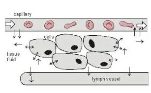
Lymph is a transudative, yellowish, and alkaline fluid. It is formed when blood plasma escapes from capillaries into the extracellular compartment (the interstitial space). While approximately 8/10 of this escaped plasma returns to the venous system, the remaining 1/10 is collected by the lymphatic system.
The composition of lymph is primarily water (96%), with the remaining 4% consisting of solutes such as proteins (2-6%), lipids (5-15%), carbohydrates, electrolytes, and cells—predominantly lymphocytes.
Diagram illustrating the exchange of substances between a capillary, tissue fluid, cells, and a lymph vessel.
Lymphatic Vessels and Drainage
The lymphatic stream follows a specific hierarchical path:
Lymphatic Capillaries: Small, blind-ended vessels that collect interstitial fluid.
Lymphatic Vessels: Larger channels that transport the fluid.
Lymph Nodes: Filtering stations where lymph is cleaned by phagocytes and macrophages.
Lymphatic Trunks: Large vessels that collect lymph from major body regions.
Collecting Ducts: The final large vessels that return lymph to the venous system.
There are two main collecting ducts: the Right Lymphatic Duct and the Thoracic Duct. The thoracic duct is the larger of the two and drains the majority of the body, while the right lymphatic duct drains specific areas on the right side of the body.
Clinical Note: Lymphoedema and Metastasis
Because the lymphatic system acts as a conduit, it can be a pathway for the spread of infections and malignant diseases (cancer). A blockage in these vessels can lead to a condition known as lymphoedema. In oncology, the "sentinel lymph node" is identified as the first node likely to receive drainage from a tumor, making it a critical site for analyzing cancer dissemination.
Organization of Lymphoid Organs
Lymphoid organs are categorized based on their role in the life cycle of a lymphocyte: whether they are "schools" for learning and maturation or "battlefields" for active defense.
Primary Lymphoid Organs
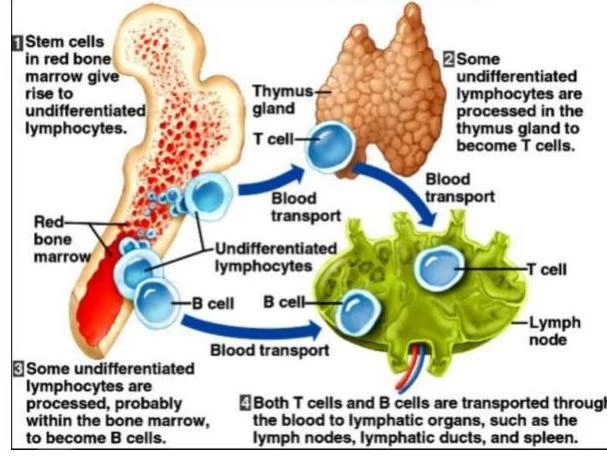
Primary lymphoid organs are the sites of hematopoiesis (the generation of blood cells) and the subsequent maturation of lymphocytes.
Bone Marrow: The origin of all blood cells via hematopoietic stem cells. It is specifically the site where B cells undergo maturation.
Thymus: A specialized organ located in the superior mediastinum, anterior to the great vessels. The thymus is essential for the maturation of T cells.
The Thymus: Development and Hormones
The thymus undergoes a distinct life cycle. It is most prominent in infants, reaching its peak size around age 2, and continues to grow until puberty. After puberty, it undergoes involution, a process where the functional thymic tissue is gradually replaced by adipose (fat) tissue.
To facilitate T cell development, the thymus secretes several critical hormones:
Thymosin: Stimulates the development of T cells.
Thymopoetin and Thymulin: Enable the distinction of T lymphocytes and enhance their functions.
This diagram illustrates the development and transport of T cells and B cells, originating from stem cells in the bone marrow and maturing in the thymus and bone marrow respectively, before circulating to lymphatic organs.
Secondary Lymphoid Organs
Secondary lymphoid organs are the sites where mature lymphocytes migrate to encounter antigens, reproduce, and differentiate into immunologically competent, antibody-secreting cells. These organs facilitate both specific and non-specific immunity and are stationed throughout the body.
Key secondary organs include:
Lymph Nodes: Kidney-shaped structures that filter lymph via afferent and efferent channels.
Spleen: An organ that filters the blood, removing dead or dying red blood cells and elaborating lymphocytes.
MALT (Mucosa-Associated Lymphoid Tissue): Includes specialized tissues like the tonsils and the aggregated lymphoid nodules (such as Peyer's patches) found in the small intestine and appendix.
Summary of Lymphatic Drainage Patterns
The drainage of lymph is organized into two distinct systems based on anatomical location:
Superficial Drainage: Vessels in the skin follow the venous drainage patterns.
Deep Drainage: Vessels in deep structures (like visceral organs) tend to accompany the arteries.
In the head and neck, these two systems are separated by the investing layer of deep fascia. Tissues superficial to this layer have their own drainage, while tissues deep to it drain into different regional and terminal lymph nodes.
Keep reading for free.
Create an account to finish this reading and make it part of your study routine.