The Lymphatic and Immune Systems: Structure and Function
An overview of the lymphatic system's roles in fluid balance, fat absorption, and immune support, alongside a detailed explanation of immune mechanisms, cellular defense, and inflammation.
The human body relies on a sophisticated, integrated network to maintain internal stability and defend against constant biological threats. This is achieved through the synergy of the lymphatic system and the immune system. While the cardiovascular system operates as a closed loop to transport blood, the lymphatic system acts as an open, unidirectional drainage system. Together, they manage fluid levels, absorb essential nutrients, and provide a specialized environment for the body's white blood cells to identify and destroy pathogens.
Functions of the Lymphatic System
The lymphatic system serves three primary roles that are essential for homeostasis:
Regulation of Interstitial Fluid Volume: As blood flows through capillaries, net filtration pressure causes water and solutes to leak into the surrounding tissue spaces, becoming interstitial fluid. If this fluid were not returned to the blood, blood volume and pressure would drop dangerously low. The lymphatic system picks up this excess fluid—at which point it is called lymph—and returns it to the cardiovascular system.
Absorption of Dietary Fats: In the small intestine, breakdown products of fats are too large to enter blood capillaries. Instead, they enter specialized lymphatic capillaries called lacteals, which transport these lipids through the lymphatic vessels to be delivered into the bloodstream.
Immune Functions: The system provides a "workspace" for the immune system. Lymphoid organs filter pathogens from the lymph and blood and serve as sites for the maturation of various leukocytes (white blood cells).
Clinical Note: Lymphedema
When lymphatic vessels are removed during surgery or blocked by pathogens (such as parasites), the return of interstitial fluid is prevented. This leads to lymphedema, a severe and often disfiguring swelling caused by the accumulation of fluid in the tissues.
Lymphatic Vessels and Circulation
Unlike the heart-driven cardiovascular system, the lymphatic system is a low-pressure, one-way circuit. Because there is no central pump, lymph flow is driven by the contraction of smooth muscle in the vessel walls and the movement of skeletal muscles.
From Capillaries to Ducts
The journey of lymph follows a specific hierarchy:
Lymphatic Capillaries: These are blind-ended tubes that surround blood capillary beds. Their endothelial cells have "flaps" that open when interstitial fluid pressure increases, allowing fluid, proteins, and even large cells like macrophages to enter.
Lymphatic Trunks: Collecting vessels merge into larger lymph trunks. There are nine major trunks, including the lumbar trunks (lower limbs/pelvis), jugular trunks (head/neck), intestinal trunk (small intestine), bronchomediastinal trunks (thoracic cavity), and subclavian trunks (upper limbs).
The Cisterna Chyli: The intestinal and lumbar trunks drain into this large, swollen vessel.
Lymphatic Ducts: All lymph eventually enters one of two ducts:
Thoracic Duct: The largest duct; it drains the entire lower body and the left side of the upper body into the junction of the left internal jugular and left subclavian veins.
Right Lymphatic Duct: Drains the upper right side of the body into the right subclavian vein.
Diagrams illustrating the main lymph trunks and ducts of the human body and the drainage point of the thoracic duct.
Lymphoid Tissues and Organs
The structural foundation of the lymphatic system is reticular tissue, a loose connective tissue containing thin fibers that form "nets" to trap pathogens. Within these nets, various lymphoid organs house leukocytes to perform immune surveillance.
Lymph Nodes
Lymph nodes are small, bean-shaped clusters located along lymphatic vessels. They act as filters, trapping approximately 90% of pathogens to prevent them from entering the bloodstream.
Structure: The node is divided into an outer cortex (rich in T cells) and an inner medulla (containing mature B cells and macrophages).
Spleen: The largest lymphoid organ. It contains red pulp, which houses macrophages that destroy old erythrocytes (red blood cells), and white pulp, which filters pathogens from the blood.
Thymus: Located in the superior mediastinum, the thymus is essential for the maturation of T cells. It is highly active in children but undergoes atrophy (replaced by fat) as an individual ages.
Mucosa-Associated Lymphatic Tissue (MALT)
MALT consists of loosely organized clusters of B and T cells that protect mucous membranes. Specific examples include:
Tonsils: Located around the oral and nasal cavities (pharyngeal, palatine, and lingual).
Peyer’s Patches: Aggregated nodules in the ileum (small intestine).
Appendix: A tube protruding from the large intestine that defends against bacteria in the gut.
Immune Mechanisms and Cellular Defense
Immunity is categorized into innate (nonspecific) immunity—the rapid, first-line response to any pathogen—and adaptive (specific) immunity—the slower, highly targeted response that possesses immunological memory.
Antigen Recognition: MHC Molecules
Adaptive immunity relies on the ability of cells to recognize antigens (glycoprotein markers) via Major Histocompatibility Complex (MHC) molecules:
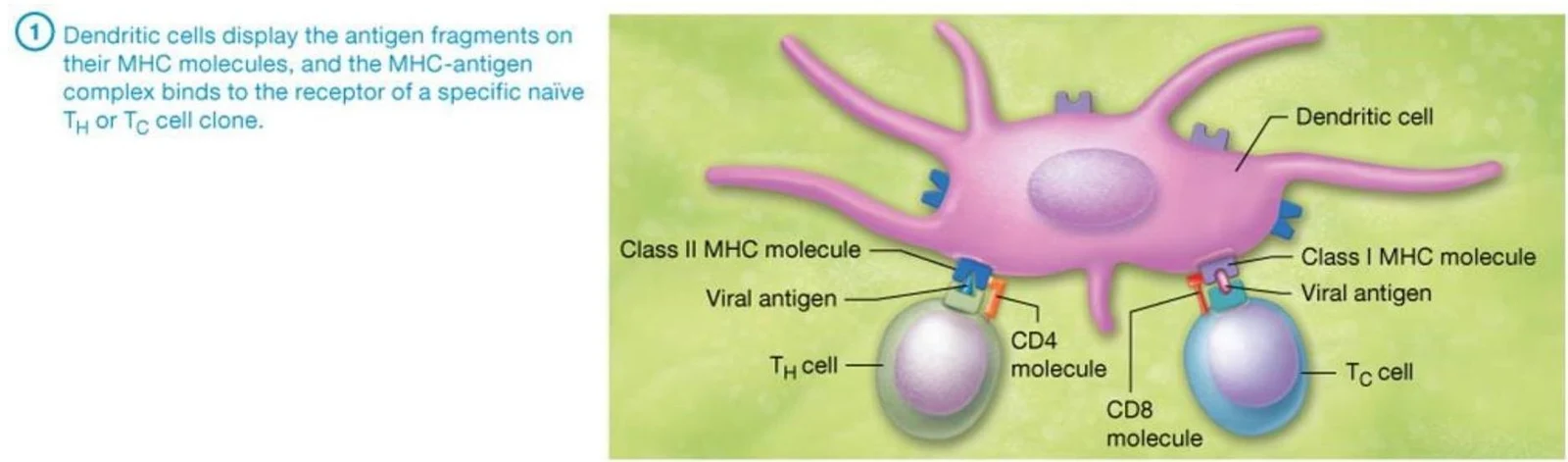
Class I MHC: Found on nearly all nucleated cells. They present endogenous antigens (synthesized inside the cell, such as viral proteins) to Cytotoxic T (TC) cells. Once activated, TC cells lyse the diseased cell.
Diagram illustrating the interaction between a naive T cell and a diseased body cell, showing the roles of the CD8 molecule, T cell receptor, endogenous antigen, and Class I MHC molecule.
Class II MHC: Found only on antigen-presenting cells (APCs), such as dendritic cells and macrophages. They present exogenous antigens (taken in via phagocytosis) to Helper T (TH) cells. Activated TH cells then act as a "warning system," secreting cytokines to stimulate other immune cells.
This illustration shows how dendritic cells present antigen fragments on MHC molecules to T cells, initiating an immune response.
Effector Mechanisms
Once a threat is identified, the body employs several strategies to eliminate it:
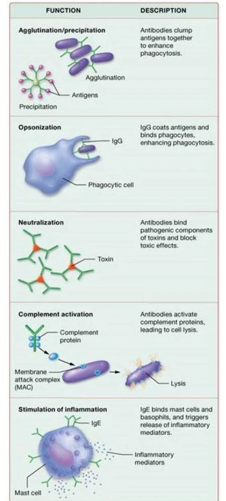
Phagocytosis & Opsonization:Macrophages and neutrophils ingest pathogens through phagocytosis. This process is greatly enhanced by opsonization, where antibodies or complement proteins coat the pathogen, making it easier for phagocytes to "grab" and ingest.
Antibody Actions: B cells produce antibodies (immunoglobulins) that fight infection through:
Agglutination: Clumping cells together.
Precipitation: Clumping soluble proteins.
Neutralization: Blocking the harmful effects of toxins or viruses.
This diagram illustrates five functions of antibodies in the immune system: agglutination/precipitation, opsonization, neutralization, complement activation, and stimulation of inflammation.
Chemical Mediators:
Pyrogens: Chemicals released by damaged cells or bacteria that act on the hypothalamus to trigger a fever.
Perforins: Secreted by T cells to create holes in the membranes of target foreign cells.
Interferons: Cytokines produced in response to viral infections that inhibit viral replication within host cells.
Inflammation: The Non-Specific Response
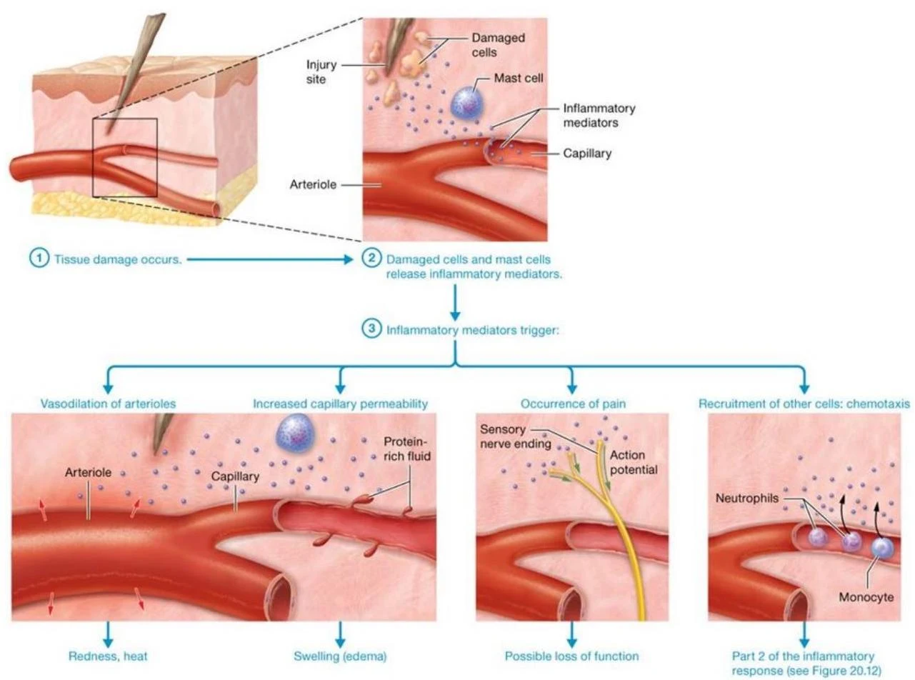
Inflammation is an innate response to cellular injury characterized by vasodilation, increased capillary permeability, pain, and phagocyte mobilization.
Stage 1: Tissue damage triggers the release of inflammatory mediators (like histamine from mast cells). This causes redness, heat, swelling (edema), and pain.
Stage 2: Phagocytes like neutrophils and macrophages are recruited to the site via chemotaxis to clean up debris and pathogens.
This diagram illustrates the process of inflammation, starting with tissue damage that triggers the release of inflammatory mediators. These mediators cause vasodilation, increased capillary permeability, pain, and the recruitment of immune cells like neutrophils and monocytes to the injury site.
Exam Tip: Comparing MHC I and MHC II
MHC I→ All nucleated cells → Presents Endogenous antigens → Activates Cytotoxic T cells.
MHC II→ APCs only → Presents Exogenous antigens → Activates Helper T cells.
Keep reading for free.
Create an account to finish this reading and make it part of your study routine.