The Hypothalamic-Pituitary Axis: Anatomy and Function
An in-depth explanation of the hypothalamic-pituitary unit, its anatomical divisions, hormonal regulation mechanisms including negative feedback and pulsatile secretion, and the roles of anterior and posterior pituitary hormones in maintaining homeostasis.
The endocrine system relies on a sophisticated hierarchy of communication to maintain homeostasis. At the center of this hierarchy is the relationship between the hypothalamus and the pituitary gland. Often referred to as the "master gland," the pituitary gland translates neural signals from the brain into hormonal messages that regulate a vast array of physiological processes, including growth, metabolism, reproduction, and water balance.
The Hypothalamic-Hypophyseal Unit
The functional connection between the brain and the endocrine system is known as the hypothalamic-hypophyseal unit. The hypothalamus acts as a command center, integrating various afferent (input) pathways from the nervous system and processing them into specific hormonal responses.
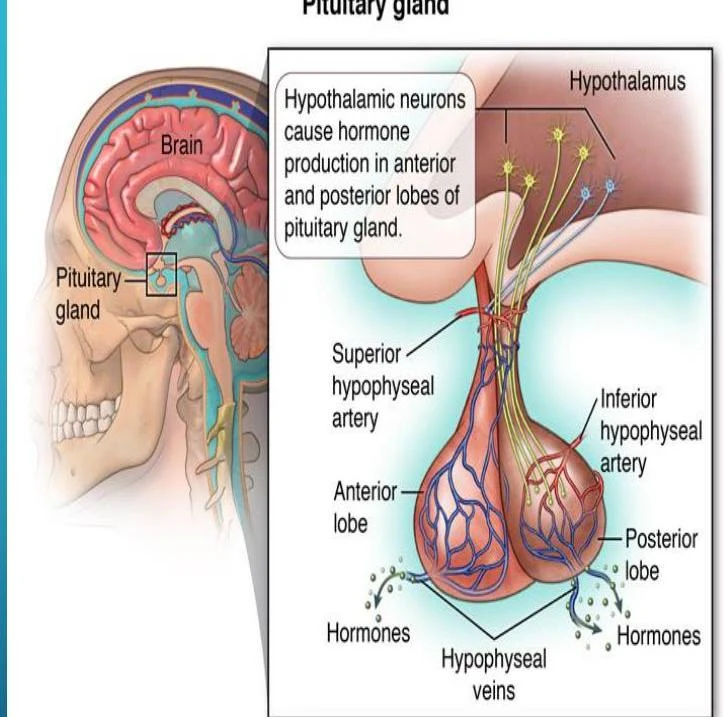
The pituitary gland itself is located within a bony pocket of the sphenoid bone and is divided into three distinct anatomical parts:
Anterior Pituitary (Adenohypophysis): The largest portion of the gland, which originates from Rathke's pouch. It receives approximately 90% of its blood supply via the hypothalamic-hypophyseal portal system, allowing hypothalamic hormones to signal the anterior lobe.
Posterior Pituitary (Neurohypophysis): An extension of the diencephalon (forebrain). Unlike the anterior lobe, it does not synthesize its own hormones; instead, it serves as a storage and release site for hormones produced in the hypothalamus.
Intermediate Lobe (Pars Intermedialis): This section is poorly developed in humans and possesses little functional capacity.
Diagram illustrating the pituitary gland and its connection to the hypothalamus, detailing the roles of hypothalamic neurons and the anterior and posterior lobes in hormone production.
Hormonal Regulation: Feedback and Secretion Patterns
The hypothalamic-hypophyseal unit does not operate at a constant level. Instead, it utilizes complex regulatory mechanisms to ensure hormone levels remain within a narrow physiological range.
Negative Feedback Mechanisms
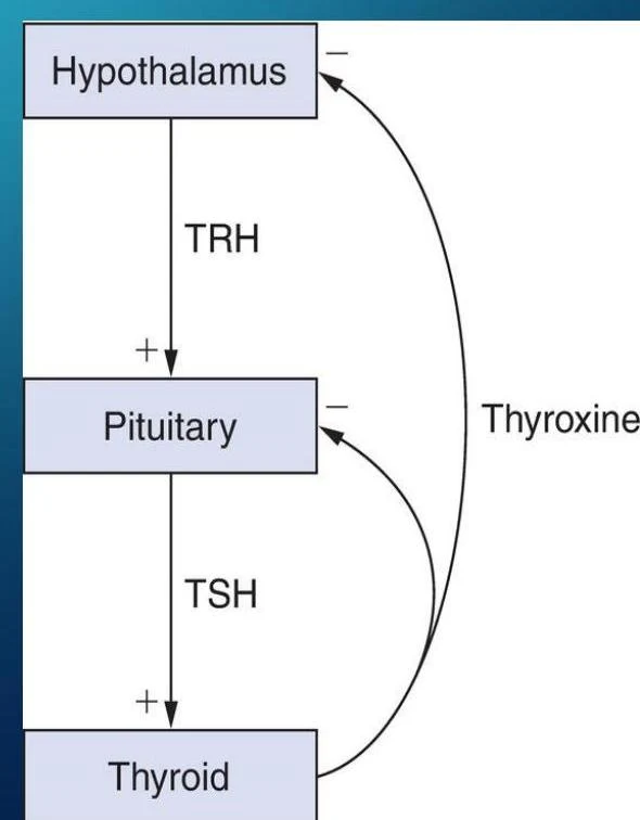
Most endocrine axes operate via "open-loop" negative feedback mechanisms. This functions much like a thermostat: when the concentration of a target hormone (the output) reaches a certain threshold, it sends a signal back to the hypothalamus and pituitary to inhibit further secretion.
This diagram illustrates the negative feedback loop of thyroid hormone regulation, involving the hypothalamus, pituitary gland, and thyroid gland.
Pulsatility and Diurnal Rhythms
To prevent receptor desensitization and allow for precise control, hormones are rarely secreted at a steady rate.
Pulsatile Secretion: All anterior pituitary hormones are secreted in pulses regulated by neural modulation.
Diurnal Rhythms (Cyclicity): The nervous system regulates cyclic functions based on external signals, such as the ratio of daylight to darkness.
Exam Tip: When interpreting lab results, remember that because of pulsatility and diurnal variation, a single measurement may not always reflect the true average hormone level of a patient.
The Anterior Pituitary (Adenohypophysis)
The anterior pituitary produces two categories of hormones: tropic hormones, which act on other endocrine glands, and direct effectors, which act directly on peripheral tissues.
Hypophysiotropic Hormones
The hypothalamus controls the anterior pituitary by secreting "releasing" or "inhibiting" hormones into the portal system.
Hormone
Action
TRH (Thyrotropin-releasing hormone)
Releases TSH and prolactin
GnRH (Gonadotropin-releasing hormone)
Releases LH and FSH
CRH (Corticotropin-releasing hormone)
Releases ACTH
GHRH (Growth hormone-releasing hormone)
Releases GH
Somatostatin
Inhibits GH and TSH release
Dopamine
Inhibits prolactin release
Key Anterior Pituitary Hormones
Growth Hormone (GH): An amphibolic hormone that influences both anabolic (building) and catabolic (breaking down) processes. It stimulates protein synthesis and the production of insulin-like growth factors (IGF-1). Excess GH leads to acromegaly in adults (characterized by bony overgrowth of the hands, feet, and face) or gigantism in children.
Prolactin (PRL): A direct effector responsible for breast development and lactation. Uniquely, prolactin is under tonic inhibition by dopamine; if dopamine is blocked or the pituitary stalk is interrupted, prolactin levels rise.
Tropic Hormones: These include TSH (targets the thyroid), ACTH (targets the adrenal cortex), and LH/FSH (target the gonads).
The Posterior Pituitary (Neurohypophysis)
The posterior pituitary does not synthesize hormones. Instead, it stores and releases two nonapeptides produced by the hypothalamus:
Arginine Vasopressin (AVP/ADH): This hormone is critical for regulating body water balance by controlling water excretion in the kidneys. A drop in blood pressure can trigger its release.
Deficiency: Leads to diabetes insipidus, characterized by polyuria (excessive urination) and polydipsia (excessive thirst).
Oxytocin: This hormone plays a vital role in reproductive processes, specifically in labor (uterine contractions) and lactation (milk let-down).
Clinical Correlations: Pituitary Dysfunction
Disruptions in this unit can lead to significant clinical pathology.
Pituitary Tumors
Pituitary adenomas are common; approximately 10-30% of healthy individuals may have them discovered incidentally during MRI scans. Functional tumors, such as prolactinomas, can cause symptoms like infertility, menstrual irregularities in women, or headaches.
Hypopituitarism
This refers to the loss of anterior pituitary function. It can be monotropic (loss of a single hormone) or panhypopituitarism (complete loss of all pituitary hormones). Causes include tumors, trauma, radiation, or infection.
Clinical Note: When evaluating suspected pituitary failure, clinicians must measure both the tropic hormone (e.g., TSH) and the target organ hormone (e.g., T4) to differentiate between primary and secondary deficiencies.
Keep reading for free.
Create an account to finish this reading and make it part of your study routine.